Human Cathepsin S Antibody
R&D Systems, part of Bio-Techne | Catalog # AF1183


Key Product Details
Validated by
Species Reactivity
Validated:
Cited:
Applications
Validated:
Cited:
Label
Antibody Source
Product Specifications
Immunogen
Gln17-Ile331
Accession # P25774
Specificity
Clonality
Host
Isotype
Scientific Data Images for Human Cathepsin S Antibody
Cathepsin S in Human Lymph Node.
Cathepsin S was detected in immersion fixed paraffin-embedded sections of human lymph node using Goat Anti-Human Cathepsin S Antigen Affinity-purified Polyclonal Antibody (Catalog # AF1183) at 15 µg/mL overnight at 4 °C. Tissue was stained using the Anti-Goat HRP-DAB Cell & Tissue Staining Kit (brown; Catalog # CTS008) and counterstained with hematoxylin (blue). View our protocol for Chromogenic IHC Staining of immersion fixed paraffin-embedded Tissue Sections.Detection of Human Cathepsin S by Western Blot
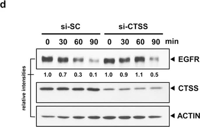
CTSS attenuates EGF-mediated EGFR degradation.(a) OEC-M1 and MDA-MB-231 cells were pretreated with 20 μM 6r or ZFL for 1 h and subsequently incubated with 100 ng/mL EGF for an additional 2 h. The total cell lysates were analysed using EGFR-specific antibodies. ACTIN was used as the internal control for semiquantitative loading in each lane. (b) The cells were stimulated with EGF (100 ng/mL) with or without the pretreatment of 20 μM 6r for the indicated durations. EGFR degradation was examined through immunostaining by using an anti-EGFR antibody. Notably, a substantial amount of EGFR was detectable even after 6 h of EGF stimulation in 6r-treated cells. (c) The OEC-M1 cells were transiently transfected with plasmids (pCMV) that encoded wild-type CTSS. After 24 h of transfection, the cells were treated with 100 ng/mL EGF for the indicated durations and the cellular EGFR, CTSS, and ACTIN signals were determined through Western blotting. The lifespan of EGF-mediated EGFR degradation was calculated by normalising the signal intensity of EGFR with that of ACTIN. (d) The MDA-MB-231 cells were transfected with specific 50 nM CTSS siRNA (si-CTSS) for 24 h and subsequently incubated with 100 ng/mL EGF for the indicated durations. The nontargeting scramble siRNA (si-SC) was used as the scramble control. (e) The MDA-MB-231 cells were transiently transfected with plasmids encoding the CTSS-C25A mutant. After 24 h of transfection, the cells were incubated with 100 ng/mL of EGF for the indicated durations. Furthermore, the cells were harvested and subjected to SDS-PAGE and Western blotting. EGFR degradation was determined using an antibody against EGFR. ACTIN signalling was included as the loading control. Image collected and cropped by CiteAb from the following open publication (https://www.nature.com/articles/srep29256), licensed under a CC-BY license. Not internally tested by R&D Systems.Detection of Human Cathepsin S by Western Blot
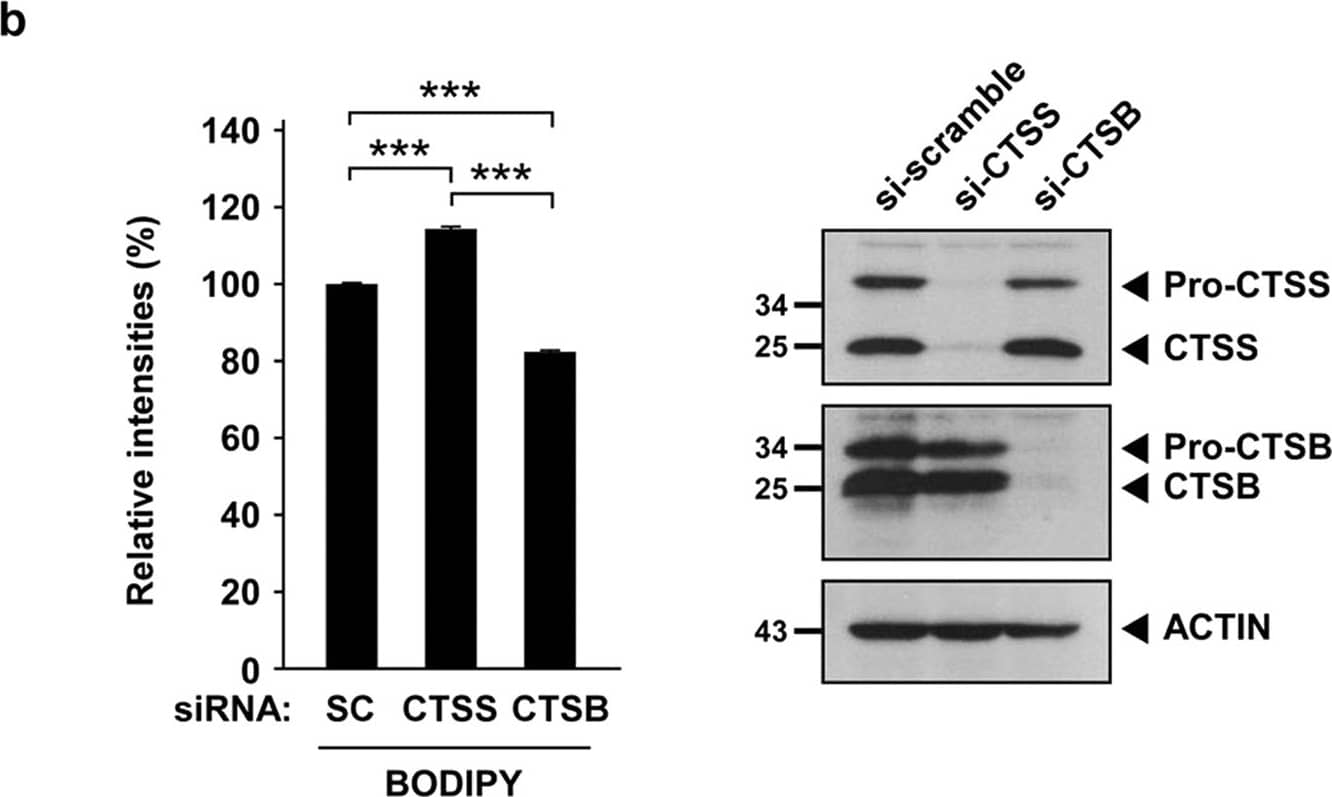
CTSS inhibition does not impair lysosomal activities.(a) OEC-M1 cells were pretreated with vesicle or 100 nM BAF for 1 h and then incubated with 20 μM 6r for 1 h further. Lysosomal proteolytic activities were determined using BODIPY–BSA and quantified through flow cytometry. (b) After 48 h of siRNA knockdown of CTSS and CTSB, the relative expression of CTSS and CTSB were determined through Western blotting (right panel). Lysosomal proteolytic activities were determined using BODIPY–BSA and quantified through flow cytometry (right panel). Data represent the mean ± SD of three independent experiments. Differences were found to be statistically significant at ***P < 0.001. Image collected and cropped by CiteAb from the following open publication (https://www.nature.com/articles/srep29256), licensed under a CC-BY license. Not internally tested by R&D Systems.Applications for Human Cathepsin S Antibody
Immunohistochemistry
Sample: Immersion fixed paraffin-embedded sections of human lymph node
Immunoprecipitation
Sample: Conditioned cell culture medium spiked with Recombinant Human Cathepsin S (Catalog # 1183-CY), see our available Western blot detection antibodies
Western Blot
Sample: Recombinant Human Cathepsin S (Catalog # 1183-CY)
Neutralization
Human Cathepsin S Sandwich Immunoassay
Reviewed Applications
Read 2 reviews rated 5 using AF1183 in the following applications:
Formulation, Preparation, and Storage
Purification
Reconstitution
Formulation
Shipping
Stability & Storage
- 12 months from date of receipt, -20 to -70 °C as supplied.
- 1 month, 2 to 8 °C under sterile conditions after reconstitution.
- 6 months, -20 to -70 °C under sterile conditions after reconstitution.
Background: Cathepsin S
Cathepsin S is a lysosomal cysteine protease of the papain family (1). It plays a major role in the processing of the MHC class II-associated invariant chain (2). It has been implicated in the pathogenesis of several diseases such as Alzheimer’s disease and degenerative disorders associated with the cells of the mononuclear phagocytic system (1). Human Cathepsin S is synthesized as a preproenzyme of 331 amino acid residues consisting a signal peptide (residues 1-16), a pro region (residues 17-114), and the mature enzyme (residues 115-331) (3-5). Cathepsin S is less abundant in tissues than Cathepsins B, L and H. The highest levels have been found in lymph nodes, spleen, macrophages, and other phagocytic cells.
References
- Kirschke, H. (2004) in Handbook of Proteolytic Enzymes (ed. Barrett, A.J. et al.) pp. 1104 - 1107, Academic Press, San Diego.
- Turk, V. et al. (2001) EMBO J. 20:4629.
- Shi, G.P. et al. (1992) J. Biol. Chem. 267:7258.
- Shi, G.P. et al. (1994) J. Biol. Chem. 269:11530.
- Wiederanders, B. et al. (1992) J. Biol. Chem. 267:13708.
Alternate Names
Entrez Gene IDs
Gene Symbol
UniProt
Additional Cathepsin S Products
Product Documents for Human Cathepsin S Antibody
Product Specific Notices for Human Cathepsin S Antibody
For research use only
