Human CD19 Antibody
R&D Systems, part of Bio-Techne | Catalog # MAB4867


Key Product Details
Species Reactivity
Validated:
Cited:
Applications
Validated:
Cited:
Label
Antibody Source
Product Specifications
Immunogen
Specificity
Clonality
Host
Isotype
Scientific Data Images for Human CD19 Antibody
Detection of CD19 in Human PBMCs by Flow Cytometry.
Human peripheral blood mononuclear cells (PBMCs) were stained with Mouse Anti-Human CD3e PE-conjugated Monoclonal Antibody (Catalog # FAB100P) and either (A) Mouse Anti-Human CD19 Monoclonal Antibody (Catalog # MAB4867) or (B) Mouse IgG1Isotype Control (Catalog # MAB002) followed by Allophycocyanin-conjugated Anti-Mouse IgG Secondary Antibody (Catalog # F0101B).Detection of Human CD19 by Flow Cytometry
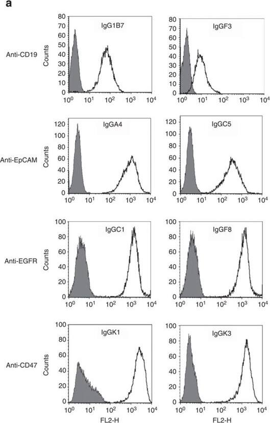
Common heavy-chain antibodies are specific and have neutralization potential.(a) Flow cytometry profiles obtained with different IgGs selected against CD19, EpCAM, EGFR and CD47. The profiles on antigen-negative versus -positive cell lines are shown shaded and open, respectively (for CD19: Jurkat/Raji; EpCAM: HEK-293/MCF-7; EGFR: MCF-7/A431; CD47: CHO/CHO-CD47). (b) Dose–response inhibition of SIRP alpha binding to CD47 expressed on cells for the positive control mAb B6H12 (open circles), IgGK1 (closed circles) and IgGK2 (crosses) that were directly isolated from the fixed heavy-chain libraries. (c) Neutralization potential in the SIRP alpha/CD47-binding assay for variants IgGK3 and IgGK4 (closed and open squares, respectively) obtained via optimization of IgGK2 (crosses). (d) Neutralization activity in the MSLN/MUC16 interaction assay for IgGO1 (open circles) and IgGO3 (closed triangles) in comparison with a negative control mAb (closed circles) and the therapeutic antibody Amatuximab (closed squares). (e) Dose–response binding profile measured by flow cytometry on CD19-positive Raji cells for IgG1B7 (open circles) and variants IgGL7-1 (triangles), and IgGL7-2 (closed circles) that were generated by affinity maturation. Error bars in b, c and d represent s.e.m. of two replicates. The data are representative of three independent experiments. Image collected and cropped by CiteAb from the following publication (https://pubmed.ncbi.nlm.nih.gov/25672245), licensed under a CC-BY license. Not internally tested by R&D Systems.Detection of Human CD19 by Flow Cytometry
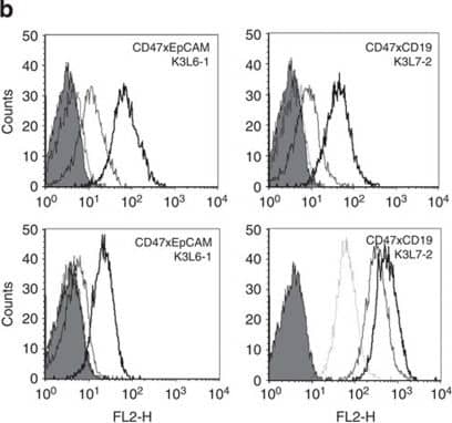
Biological activity of bispecific kappa lambda-bodies.(a) Schematic representation of monovalent and bivalent binding of CD19xCD47 kappa lambda-bodies on the surface of CD47+/CD19−DS-1 cells (top) and CD47+/CD19+Raji cells (bottom). (b) Binding of CD47 × EpCAM (left panels) and CD47 × CD19 kappa lambda-bodies (right panels) to DS-1 (top panels) and Raji cells (bottom panels) monitored by FACS. kappa lambda bodies were used at 0.01, 0.1 and 1 mg ml−1, control staining with fluorescently labelled secondary antibody is indicated by the shaded area. (c) Inhibition of SIRP alpha binding to the CD47 on DS-1 (top) and Raji cells (bottom) by increasing concentrations of different antibodies: anti-CD47-positive control mAb B6H12 (grey open circles); CD47 × CD19 K3L7-1 (black open circles, dotted line); CD47 × CD19 K3L7-2 (closed circles); CD47 × EpCAM K3L6-1 (open triangles). Error bars represent s.e.m. of four replicates and the data are representative of three independent experiments. (d) T-cell-mediated killing of EpCAM-positive cells by CD3 × EpCAM kappa lambda-bodies. MCF-7 cells were incubated with purified human T cells in the presence of increasing concentrations of different antibodies. CD3 × EpCAM kappa lambda-bodies: L6-3L3-1 (closed circles), L6-4L3-1 (closed squares), L6-5L3-1 (closed triangles); anti-EpCAM mAbs: L6-3 (open circles), L6-4 (open squares), L6-5 (open triangles); anti-CD3 mAb L3-1 (crosses). Error bars represent s.d. of three replicates and the experiment was repeated with the blood of two independent donors. The isoelectric focusing (IEF) gels and electrophoresis profiles corresponding to the different kappa lambda-bodies and mAbs are shown in Fig. 3 and Supplementary Fig. 1. Image collected and cropped by CiteAb from the following publication (https://pubmed.ncbi.nlm.nih.gov/25672245), licensed under a CC-BY license. Not internally tested by R&D Systems.Applications for Human CD19 Antibody
CyTOF-ready
Flow Cytometry
Sample: Human peripheral blood mononuclear cells (PBMCs)
Reviewed Applications
Read 1 review rated 4 using MAB4867 in the following applications:
Formulation, Preparation, and Storage
Purification
Reconstitution
Formulation
Shipping
Stability & Storage
- 12 months from date of receipt, -20 to -70 °C as supplied.
- 1 month, 2 to 8 °C under sterile conditions after reconstitution.
- 6 months, -20 to -70 °C under sterile conditions after reconstitution.
Background: CD19
CD19 is a 95 kDa transmembrane glycoprotein with two Ig-like C2-set domains. CD19 regulates B cell development and activation through interactions with CD21, CD22, and the B cell receptor. CD19 polymorphisms and up‑regulation lead to the development of autoimmunity by promoting autoantibody production. Within the extracellular domain, human CD19 (Accession # P15391) shares 57% amino acid sequence identity with mouse and rat CD19.
Alternate Names
Entrez Gene IDs
Gene Symbol
Additional CD19 Products
Product Documents for Human CD19 Antibody
Product Specific Notices for Human CD19 Antibody
For research use only

