Human CD34 APC-conjugated Antibody
R&D Systems, part of Bio-Techne | Catalog # FAB7227A


Conjugate
Catalog #
Key Product Details
Species Reactivity
Validated:
Human
Cited:
Human, Mouse
Applications
Validated:
Flow Cytometry
Cited:
CAR-T (Flow Cytometry), Flow Cytometry, Immunocytochemistry
Label
Allophycocyanin (Excitation = 620-650 nm, Emission = 660-670 nm)
Antibody Source
Monoclonal Mouse IgG1 Clone # QBEnd10
Product Specifications
Immunogen
Human endothelial vesicles
Specificity
Detects human CD34 in direct ELISAs.
Clonality
Monoclonal
Host
Mouse
Isotype
IgG1
Scientific Data Images for Human CD34 APC-conjugated Antibody
Detection of CD34 in Human PBMCs by Flow Cytometry.
Human peripheral blood mononuclear cells (PBMCs) were stained with Mouse Anti-Human CD34 APC-conjugated Monoclonal Antibody (Catalog # FAB7227A) and Mouse Anti-Human CD45 PE-conjugated Monoclonal Antibody (Catalog # FAB1430P). Quadrant markers were set based on control antibody staining (Catalog # IC002A). View our protocol for Staining Membrane-associated Proteins.Detection of CD34 in KG‑1a Human Cell Line by Flow Cytometry.
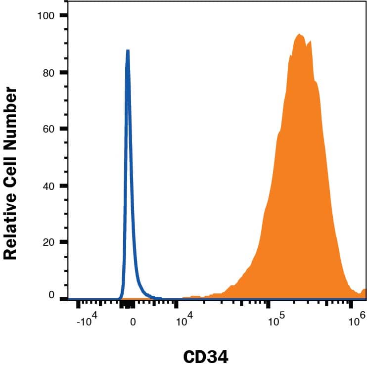
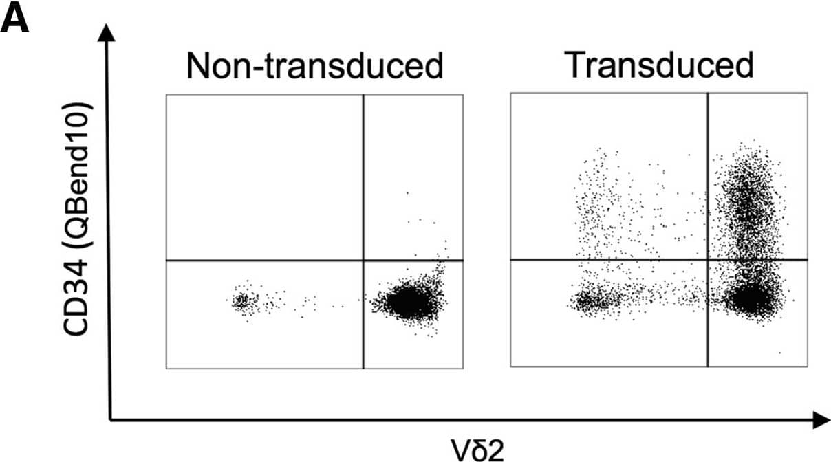
KG-1a human acute myelogenous leukemia cell line was stained with Mouse Anti-Human CD34 APC-conjugated Monoclonal Antibody (Catalog # FAB7227A, filled histogram) or isotype control antibody (Catalog # IC002A, open histogram). View our protocol for Staining Membrane-associated Proteins.Detection of Human CD34 by Flow Cytometry
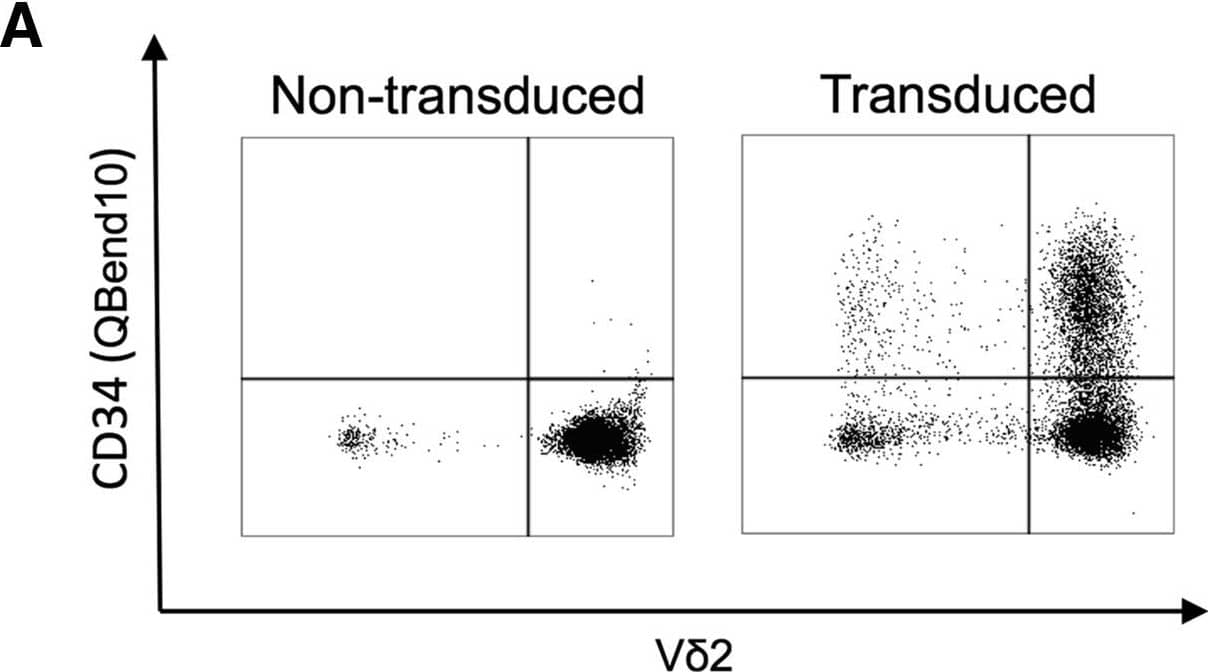
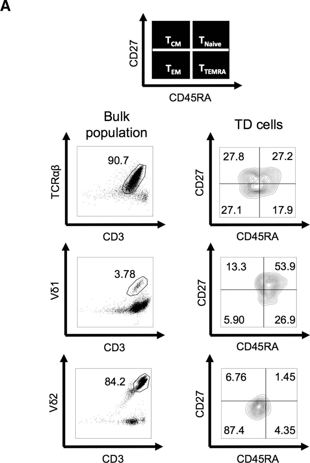
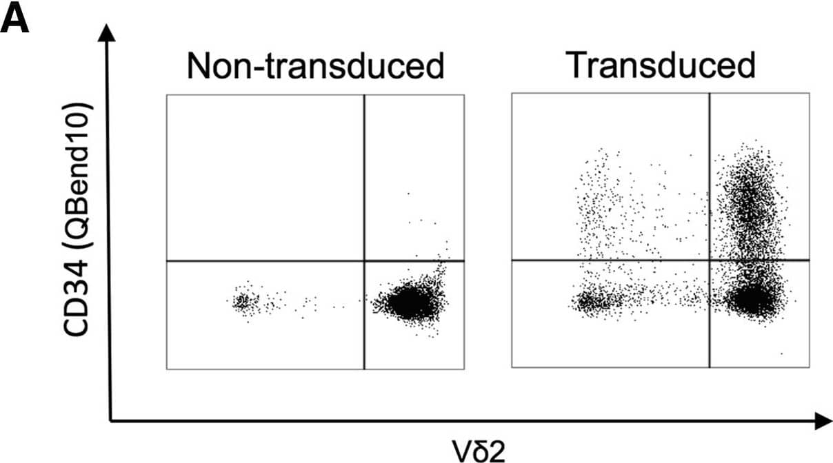
alpha beta, V delta1+, and V delta2+ T Cells Are Efficiently Transduced with GD2-CAR following Activation with CD3/CD28 Antibody, ZOL, or ConA, and Bulk Populations Are Cytotoxic to Neuroblastoma Cells(A) Representative flow cytometry dot plot showing transduction efficiency of ZOL-expanded non-transduced and GD2-CAR+-transduced PBMCs. V delta2+ populations were gated on CD3+ live cells 8 days following transduction. The GD2-CAR construct coexpresses the QBend10 epitope from CD34, allowing detection by flow cytometry. Transduction efficiency was determined by the percentage of QBend10+ in the T cell population gate compared to control non-transduced cells. (B–D) Mean transduction efficiency using CD3/CD28 antibody, ZOL, or ConA activation methods, respectively. Each data point represents an individual donor (n = 9) and each horizontal line is the mean. (E–G) Bulk populations of GD2-CAR-transduced T cells stimulated by CD3/CD28 antibody (E), ZOL (F), and ConA (G) specifically lyse the GD2-expressing neuroblastoma cell line, LAN1, in 4-hr 51Cr release assay. NTD, non-transduced T cells; TD, transduced GD2-CAR+ T cells (data represented as mean ± SEM; 3–5 individual donors in triplicate). Image collected and cropped by CiteAb from the following publication (https://linkinghub.elsevier.com/retrieve/pii/S1525001617305981), licensed under a CC-BY license. Not internally tested by R&D Systems.Applications for Human CD34 APC-conjugated Antibody
Application
Recommended Usage
Flow Cytometry
10 µL/106 cells
Sample: Human peripheral blood mononuclear cells (PBMCs) and KG‑1a human acute myelogenous leukemia cell line
Sample: Human peripheral blood mononuclear cells (PBMCs) and KG‑1a human acute myelogenous leukemia cell line
Formulation, Preparation, and Storage
Purification
Protein A or G purified from hybridoma culture supernatant
Formulation
Supplied in a saline solution containing BSA and Sodium Azide.
Shipping
The product is shipped with polar packs. Upon receipt, store it immediately at the temperature recommended below.
Stability & Storage
Protect from light. Do not freeze.
- 12 months from date of receipt, 2 to 8 °C as supplied.
Background: CD34
Alternate Names
CD34, HPCA1
Gene Symbol
CD34
Additional CD34 Products
Product Documents for Human CD34 APC-conjugated Antibody
Product Specific Notices for Human CD34 APC-conjugated Antibody
For research use only
Loading...
Loading...
Loading...
Loading...
Loading...
Loading...
