Human CIS-1 Antibody
R&D Systems, part of Bio-Techne | Catalog # AF3194

Key Product Details
Species Reactivity
Validated:
Cited:
Applications
Validated:
Cited:
Label
Antibody Source
Product Specifications
Immunogen
Leu11-Leu258
Accession # Q9NSE2
Specificity
Clonality
Host
Isotype
Scientific Data Images for Human CIS-1 Antibody
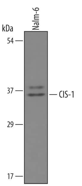
Detection of Human CIS-1 by Western Blot.
Western blot shows lysates of Nalm-6 human Pre-B acute lymphocytic leukemia cell line. PVDF membrane was probed with 1 µg/mL of Goat Anti-Human CIS-1 Antigen Affinity-purified Polyclonal Antibody (Catalog # AF3194) followed by HRP-conjugated Anti-Goat IgG Secondary Antibody (Catalog # HAF019). A specific band was detected for CIS-1 at approximately 35-40 kDa (as indicated). This experiment was conducted under reducing conditions and using Immunoblot Buffer Group 8.CIS-1 in HDLM-2 Human Cell Line.
CIS-1 was detected in immersion fixed HDLM-2 human Hodgkin’s lymphoma cell line using Goat Anti-Human CIS-1 Antigen Affinity-purified Polyclonal Antibody (Catalog # AF3194) at 15 µg/mL for 3 hours at room temperature. Cells were stained using the NorthernLights™ 557-conjugated Anti-Goat IgG Secondary Antibody (red; NL001) and counterstained with DAPI (blue). Specific staining was localized to cytoplasm. Staining was performed using our protocol for Fluorescent ICC Staining of Non-adherent Cells.Detection of Human CIS-1 by Simple WesternTM.
Simple Western lane view shows lysates of NK human natural killer lymphoma cell line, loaded at 0.2 mg/mL. Specific bands were detected for CIS-1 at approximately 37 and 42 kDa (as indicated) using 10 µg/mL of Goat Anti-Human CIS-1 Antigen Affinity-purified Polyclonal Antibody (Catalog # AF3194) . This experiment was conducted under reducing conditions and using the 12-230 kDa separation system.Applications for Human CIS-1 Antibody
Immunocytochemistry
Sample: Immersion fixed HDLM‑2 human Hodgkin's lymphoma cell line
Simple Western
Sample: NK human natural killer lymphoma cell line
Western Blot
Sample: Nalm-6 human Pre-B acute lymphocytic leukemia cell line
Reviewed Applications
Read 1 review rated 1 using AF3194 in the following applications:
Formulation, Preparation, and Storage
Purification
Reconstitution
Formulation
Shipping
Stability & Storage
- 12 months from date of receipt, -20 to -70 °C as supplied.
- 1 month, 2 to 8 °C under sterile conditions after reconstitution.
- 6 months, -20 to -70 °C under sterile conditions after reconstitution.
Background: CIS-1
Cytokine Inducible SH2-containing protein (CIS-1) is a 29 kDa protein found in a variety of cell types. Mono or polyubiquitination generally results in a 37 or 45 kDa molecule. CIS-1 binds to phosphorylated cytokine receptors IL-3 R beta and EPO-R and blocks downstream activation of STAT5 via receptor internalization and ubiquitin‑mediated proteosomal degradation. Human CIS-1 is a 258 aa peptide that contains one SH2 domain (aa 82‑163) and one SOCS box (aa 218‑258). There are two known alternatively spliced variants with a 7- or 13-aa substitution for the 7 N-terminal amino acid residues. Over the region used as immunogen, human CIS-1 is 91% identical to the corresponding mouse and canine protein sequences.
Long Name
Alternate Names
Gene Symbol
UniProt
Additional CIS-1 Products
Product Documents for Human CIS-1 Antibody
Product Specific Notices for Human CIS-1 Antibody
For research use only


