Human CL-P1/COLEC12 Antibody
R&D Systems, part of Bio-Techne | Catalog # AF2690

Key Product Details
Species Reactivity
Validated:
Cited:
Applications
Validated:
Cited:
Label
Antibody Source
Product Specifications
Immunogen
Leu57-Leu742
Accession # Q5KU26
Specificity
Clonality
Host
Isotype
Scientific Data Images for Human CL-P1/COLEC12 Antibody
Detection of CL‑P1/COLEC12 in HUVEC Human Cells by Flow Cytometry.
HUVEC human umbilical vein endothelial cells were stained with Goat Anti-Human CL-P1/COLEC12 Antigen Affinity-purified Polyclonal Antibody (Catalog # AF2690, filled histogram) or isotype control antibody (Catalog # AB-108-C, open histogram), followed by Allophycocyanin-conjugated Anti-Goat IgG Secondary Antibody (Catalog # F0108).Detection of Human CL-P1/COLEC12 by Immunocytochemistry/Immunofluorescence
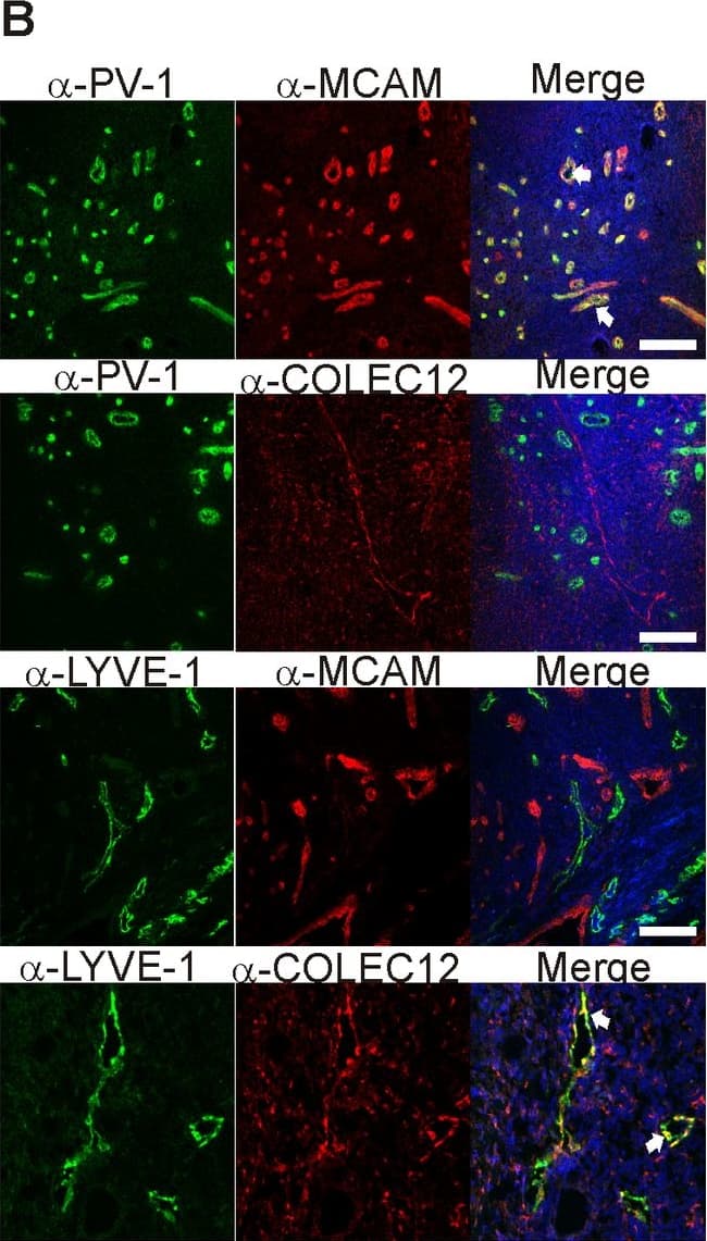
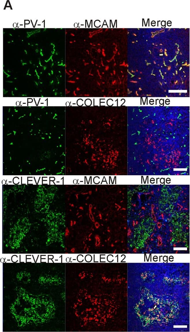
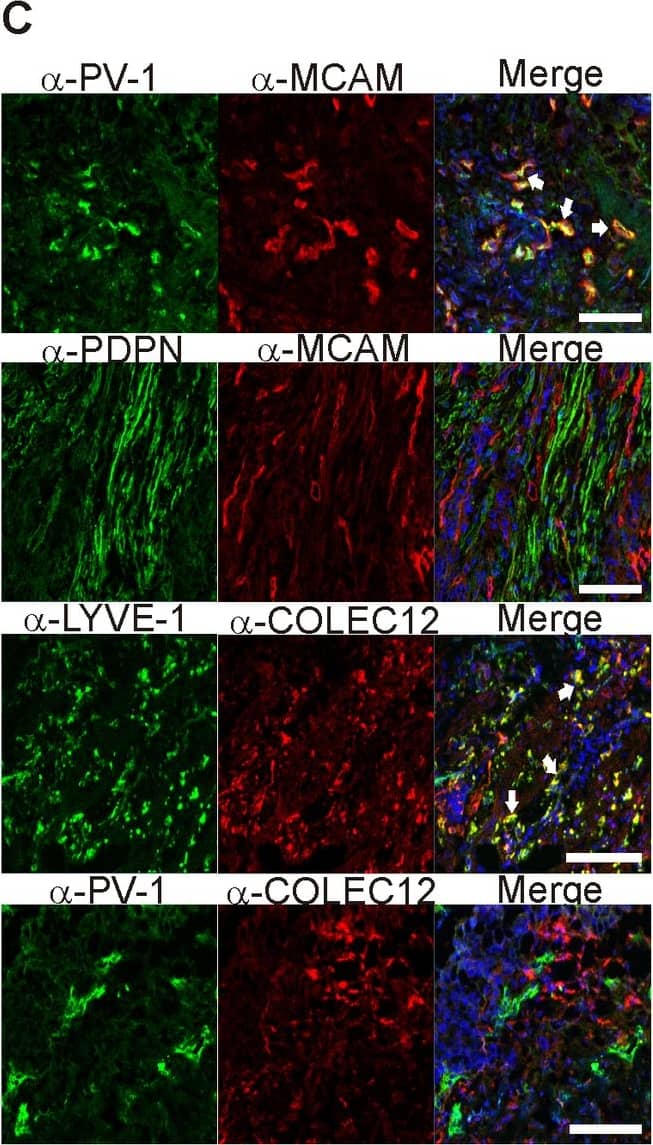
MCAM and COLEC12 are novel BEC- and LEC- specific markers.Two new endothelial markers MCAM and COLEC12 (selected from our analysis of Dataset B) were used to stain (A) normal human lymph nodes together with two established vascular markers PV-1 (for BEC) and CLEVER-1 (for LEC). In addition, (B) chronically inflamed tonsils and (C) specimens from bladder cancer and colorectal cancer were stained with the antibodies against the indicated proteins. LYVE-1 and COLEC12 co-staining was done on colorectal cancer specimens, whereas the other stainings represent bladder cancer. MCAM staining colocalized very well with the established BEC marker PV-1 whereas no colocalization could be detected between MCAM and the established LEC markers LYVE-1 and podoplanin. COLEC12-staining on the other hand showed colocalization with LYVE-1 but not PV-1. White arrows point to areas of colocalization. Nuclear counterstaining was performed with DAPI. Scale bars represent 100 µm. Image collected and cropped by CiteAb from the following publication (https://pubmed.ncbi.nlm.nih.gov/24058540), licensed under a CC-BY license. Not internally tested by R&D Systems.Detection of Human CL-P1/COLEC12 by Immunocytochemistry/Immunofluorescence
MCAM and COLEC12 are novel BEC- and LEC- specific markers.Two new endothelial markers MCAM and COLEC12 (selected from our analysis of Dataset B) were used to stain (A) normal human lymph nodes together with two established vascular markers PV-1 (for BEC) and CLEVER-1 (for LEC). In addition, (B) chronically inflamed tonsils and (C) specimens from bladder cancer and colorectal cancer were stained with the antibodies against the indicated proteins. LYVE-1 and COLEC12 co-staining was done on colorectal cancer specimens, whereas the other stainings represent bladder cancer. MCAM staining colocalized very well with the established BEC marker PV-1 whereas no colocalization could be detected between MCAM and the established LEC markers LYVE-1 and podoplanin. COLEC12-staining on the other hand showed colocalization with LYVE-1 but not PV-1. White arrows point to areas of colocalization. Nuclear counterstaining was performed with DAPI. Scale bars represent 100 µm. Image collected and cropped by CiteAb from the following publication (https://pubmed.ncbi.nlm.nih.gov/24058540), licensed under a CC-BY license. Not internally tested by R&D Systems.Applications for Human CL-P1/COLEC12 Antibody
Blockade of Receptor-ligand Interaction
CyTOF-ready
Flow Cytometry
Sample: HUVEC human umbilical vein endothelial cells
Immunocytochemistry
Sample: Immersion fixed HUVEC human umbilical vein endothelial cells
Western Blot
Sample: Recombinant Human CL-P1/COLEC12 (Catalog # 2690-CL)
Reviewed Applications
Read 1 review rated 5 using AF2690 in the following applications:
Formulation, Preparation, and Storage
Purification
Reconstitution
Formulation
Shipping
Stability & Storage
- 12 months from date of receipt, -20 to -70 °C as supplied.
- 1 month, 2 to 8 °C under sterile conditions after reconstitution.
- 6 months, -20 to -70 °C under sterile conditions after reconstitution.
Background: CL-P1/COLEC12
Collectins are a family of Ca++-dependent, C-type lectins that contain a collagenous domain and function as recognition molecules for molecular patterns found on pathogens (1‑4). Human collectin placenta 1 (CL-P1; also known as collectin sub-family member 12 and SRCL type I [scavenger receptor with C-type lectin type I]) is a 110 kDa member of the collectin family of glycoproteins (5, 6). CL-P1 is the only collectin known to be membrane bound, while CL-L1 (collectin liver-1) is the only known cytoplasmic collectin (1). Human CL-P1 is synthesized as a 742 amino acid (aa) type II transmembrane glycoprotein that contains an N-terminal 39 aa cytoplasmic domain, a 17 aa transmembrane segment, and a 686 aa C-terminal extracellular region (6). The short cytoplasmic domain contains an internalization motif (Y-K-R-F) while the extracellular region is complex, demonstrating a coiled-coil segment, a Ser-Thr rich region, a collagen-like structure and a C-type lectin/ carbohydrate recognition domain (CRD). Notably, this CRD recognizes galactose (and fucose) within the context of asialo‑orosomucoids associated with the Lewisx epitope (7, 8). CL-P1 has a 300 kDa trimeric form due to its collagen-like and coiled-coil helical domains (1, 5). There is a 97 kDa, alternate splice form of CL-P1 (SRCL type II) that shows a 120 aa truncation at the C-terminus. This effectively removes the entire CRD found on full‑length CL-P1 (6). Human CL-P1 is 93% aa identical to mouse CL-P1 over the entire extracellular region, and 87% aa identical over the CRD region (5, 9). Human CL-P1 is known to be expressed in vascular endothelial cells (5).
References
- van de Wetering, JK. et al. (2004) Eur. J. Biochem. 271:1229.
- Holmskov, U. et al. (2003) Annu. Rev. Immunol. 21:547.
- Hoppe, H-J. and K. Reid (1994) Protein Sci. 3:1143.
- Hickling, T.P. et al. (2004) J. Leukoc. Biol. 75:27.
- Ohtani, K. et al. (2001) J. Biol. Chem. 276:44222.
- Nakamura,K. et al. (2001) Biochem. Biophys. Res. Commun. 280:1028.
- Coombs, P.J. et al. (2005) J. Biol. Chem. 280:22993.
- Yoshida, T. et al. (2003) J. Biochem. 133:271.
- Nakamura, K. et al. (2001) Biochim. Biophys. Acta 1522:53.
Long Name
Alternate Names
Gene Symbol
UniProt
Additional CL-P1/COLEC12 Products
Product Documents for Human CL-P1/COLEC12 Antibody
Product Specific Notices for Human CL-P1/COLEC12 Antibody
For research use only


