Human CXCR7/RDC-1 Antibody
R&D Systems, part of Bio-Techne | Catalog # MAB4227

Key Product Details
Validated by
Species Reactivity
Validated:
Cited:
Applications
Validated:
Cited:
Label
Antibody Source
Product Specifications
Immunogen
Met1-Lys362 (Gly131Ser)
Accession # AAA62370
Specificity
Clonality
Host
Isotype
Scientific Data Images for Human CXCR7/RDC-1 Antibody
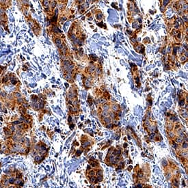
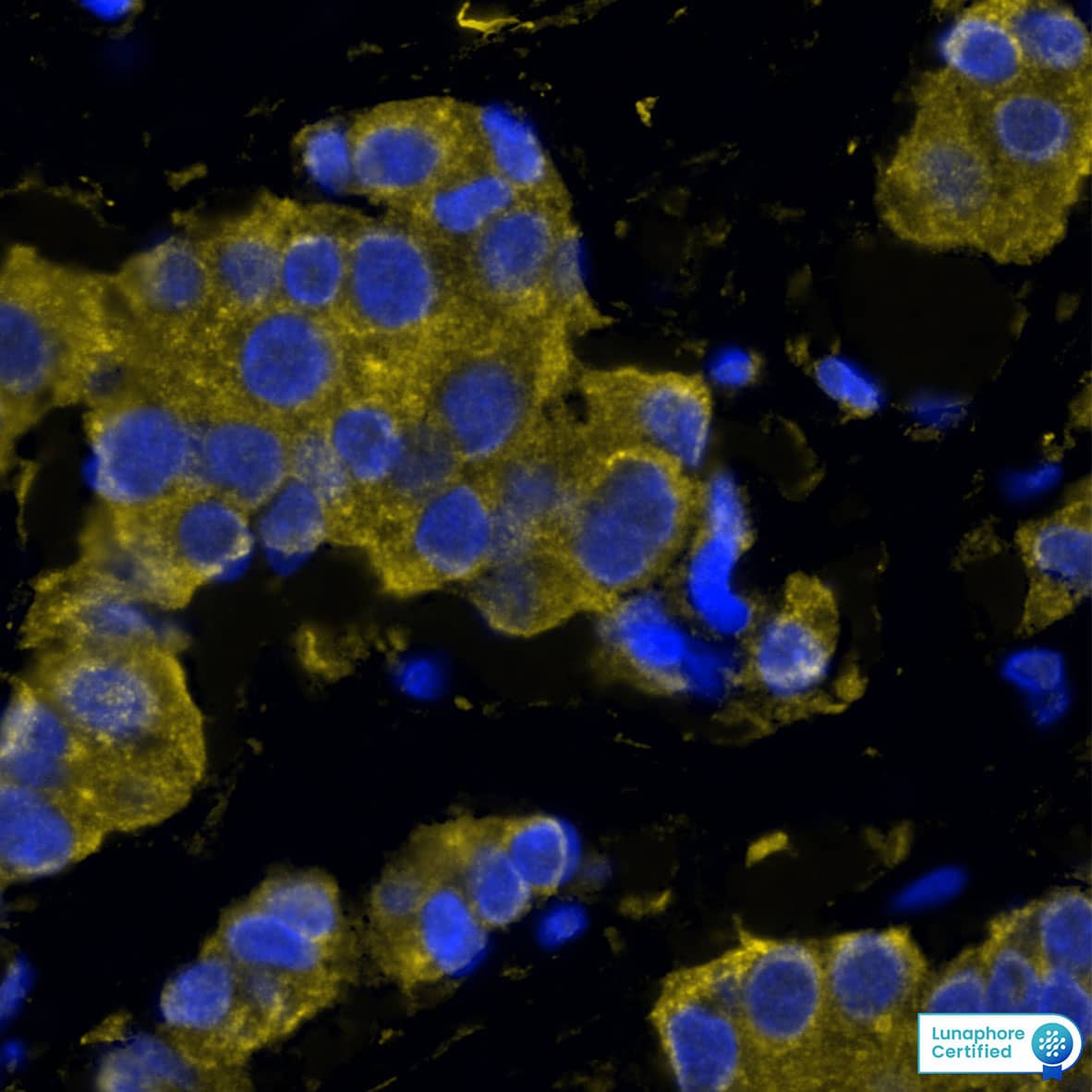
Detection of CXCR7 in Human Breast Tumor via Multiplex Immunofluorescence staining on COMET™
CXCR7 was detected in immersion fixed paraffin-embedded sections of human breast tumor using Mouse Anti-Human CXCR7 Monoclonal Antibody (Catalog # MAB4227) at 15ug/mL at 37 ° Celsius for 4 minutes. Before incubation with the primary antibody, tissue underwent an all-in-one dewaxing and antigen retrieval preprocessing using PreTreatment Module (PT Module) and Dewax and HIER Buffer H (pH 9). Tissue was stained using the Alexa Fluor™ 647 Goat anti-Mouse IgG Secondary Antibody at 1:200 at 37 ° Celsius for 2 minutes. (Yellow; Lunaphore Catalog # DR647MS) and counterstained with DAPI (blue; Lunaphore Catalog # DR100). Specific staining was localized to the cytoplasm. Protocol available in COMET™ Panel Builder.Detection of CXCR7/RDC‑1 in Human peripheral blood Monocytes by Flow Cytometry.
Human peripheral blood monocytes were stained with Mouse Anti-Human CXCR7/RDC-1 Monoclonal Antibody (Catalog # MAB4227, filled histogram) or isotype control antibody (Catalog # MAB003, open histogram), followed by Allophycocyanin-conjugated Anti-Mouse IgG F(ab')2Secondary Antibody (Catalog # F0101B).CXCR7/RDC‑1 in Human Breast Cancer Tissue.
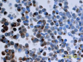
CXCR7/RDC-1 was detected in perfusion fixed paraffin-embedded sections of nude mice injected with human breast cancer cells using Mouse Anti-Human CXCR7/RDC-1 Monoclonal Antibody (Catalog # MAB4227) at 25 µg/mL overnight at 4 °C. Tissue was stained using the Anti-Mouse HRP-DAB Cell & Tissue Staining Kit (brown; Catalog # CTS002) and counterstained with hematoxylin (blue). View our protocol for Chromogenic IHC Staining of Paraffin-embedded Tissue Sections.Applications for Human CXCR7/RDC-1 Antibody
CyTOF-ready
Flow Cytometry
Sample: Human peripheral blood monocytes
Immunohistochemistry
Sample: Perfusion fixed paraffin-embedded sections of nude mice injected with human breast cancer cells and immersion fixed paraffin-embedded sections of human breast cancer tissue
Multiplex Immunofluorescence
Sample: Immersion fixed paraffin-embedded sections of Human Breast Cancer tissue
Formulation, Preparation, and Storage
Purification
Reconstitution
Formulation
*Small pack size (-SP) is supplied either lyophilized or as a 0.2 µm filtered solution in PBS.
Shipping
Stability & Storage
- 12 months from date of receipt, -20 to -70 °C as supplied.
- 1 month, 2 to 8 °C under sterile conditions after reconstitution.
- 6 months, -20 to -70 °C under sterile conditions after reconstitution.
Background: CXCR7/RDC-1
The G protein-coupled receptor, RDC1, belongs to a subgroup of chemokine receptors and has been designated CXCR7. CXCR7 can bind with high-affinity to CXCL12/SDF-1 and CXCL11/I-TAC. It is also a co-receptor for several HIV and SIV strains. In their N-termini and extracellular loops 1, 2, and 3, human and mouse CXCR7 share 84%, 100%, 96% and 86% amino acid sequence identity, respectively. Reports of mRNA levels and/or protein expression (as assessed using anti‑CXCR7, clone 9C4) (1, 2) indicate that CXCR7 occurs on a wide variety of tissues and cells including monocytes, B cells, T cells and mature dendritic cells. In contrast, based on ligand binding analysis and receptor level (as assessed using anti‑CXCR7, clone 11G8), surface expression of CXCR7 was reported to be restricted to tumor cells, activated endothelial cells, fetal liver cells, and few other cell types (3). The basis of these inconsistent observations is not known but may be attributed to cell context and the use of different antibodies that may recognize different epitopes.
References
- Balabanian, K. et al. (2005) J. Biol. Chem. 280:35760.
- Infantino, S. et al. (2006) J. Immunol. 176:2197.
- Burns, J.M. et al. (2006) J. Exp. Med. 203:2201.
Alternate Names
Gene Symbol
UniProt
Additional CXCR7/RDC-1 Products
Product Documents for Human CXCR7/RDC-1 Antibody
Product Specific Notices for Human CXCR7/RDC-1 Antibody
For research use only

