Human FABP5/E-FABP Antibody
R&D Systems, part of Bio-Techne | Catalog # MAB3077


Key Product Details
Validated by
Species Reactivity
Validated:
Cited:
Applications
Validated:
Cited:
Label
Antibody Source
Product Specifications
Immunogen
Ala2-Glu135
Accession # Q01469
Specificity
Clonality
Host
Isotype
Scientific Data Images for Human FABP5/E-FABP Antibody
Detection of Human FABP5/E‑FABP by Western Blot.
Western blot shows lysates of human heart tissue, human brain (cerebellum) tissue, and human placenta tissue. PVDF membrane was probed with 1 µg/mL of Rat Anti-Human FABP5/E-FABP Monoclonal Antibody (Catalog # MAB3077) followed by HRP-conjugated Anti-Rat IgG Secondary Antibody (Catalog # HAF005). A specific band was detected for FABP5/ E-FABP at approximately 15 kDa (as indicated). This experiment was conducted under reducing conditions and using Immunoblot Buffer Group 1.FABP5 in HUVEC Human Cells.
FABP5 was detected in immersion fixed HUVEC human umbilical vein endothelial cells using Rat Anti-Human FABP5 Monoclonal Antibody (Catalog # MAB3077) at 10 µg/mL for 3 hours at room temperature. Cells were stained using the Northern-Lights™ 557-con-jugated Anti-Rat IgG Secondary Antibody (yellow; Catalog # NL013) and counter-stained with DAPI (blue). View our protocol for Fluorescent ICC Staining of Cells on Coverslips.Western Blot Shows Human FABP5/E‑FABP Specificity by Using Knockout Cell Line.
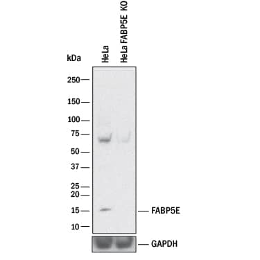
Western blot shows lysates of HeLa human cervical epithelial carcinoma parental cell line and FABP5/E-FABP knockout HeLa cell line (KO). PVDF membrane was probed with 1 µg/mL of Rat Anti-Human FABP5/E-FABP Monoclonal Antibody (Catalog # MAB3077) followed by HRP-conjugated Anti-Rat IgG Secondary Antibody (Catalog # HAF005). A specific band was detected for FABP5/E-FABP at approximately 15 kDa (as indicated) in the parental HeLa cell line, but is not detectable in knockout HeLa cell line. GAPDH (Catalog # MAB5718) is shown as a loading control. This experiment was conducted under reducing conditions and using Immunoblot Buffer Group 1.Applications for Human FABP5/E-FABP Antibody
CyTOF-ready
Flow Cytometry
Sample: HUVEC human umbilical vein endothelial cells
Immunocytochemistry
Sample: Immersion fixed HUVEC human umbilical vein endothelial cells
Knockout Validated
Western Blot
Sample: Human heart tissue, human brain (cerebellum) tissue, and human placenta tissue
Formulation, Preparation, and Storage
Purification
Reconstitution
Formulation
Shipping
Stability & Storage
- 12 months from date of receipt, -20 to -70 °C as supplied.
- 1 month, 2 to 8 °C under sterile conditions after reconstitution.
- 6 months, -20 to -70 °C under sterile conditions after reconstitution.
Background: FABP5/E-FABP
FABP5, also known as epidermal fatty acid binding protein (E-FABP), is expressed in skin, lens, adipose tissue, endothelial cells, heart, brain and placenta. FABP-5 is associated with keratinocytes and adipocytes, and is suggested to promote fatty acid availability to enzymes, protect cell structures from fatty acid attack, and target fatty acids to nuclear transcription factors. Human FABP-5 shares 80%, 81%, and 92% aa identity with mouse, rat and bovine FABP-5, respectively.
Long Name
Alternate Names
Gene Symbol
UniProt
Additional FABP5/E-FABP Products
Product Documents for Human FABP5/E-FABP Antibody
Product Specific Notices for Human FABP5/E-FABP Antibody
For research use only

