Human FAK Antibody
R&D Systems, part of Bio-Techne | Catalog # MAB4467

Key Product Details
Validated by
Species Reactivity
Validated:
Cited:
Applications
Validated:
Cited:
Label
Antibody Source
Product Specifications
Immunogen
Asp213-Thr412
Accession # Q05397
Specificity
Clonality
Host
Isotype
Scientific Data Images for Human FAK Antibody
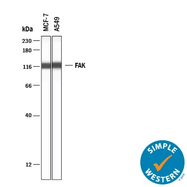
Detection of Human FAK by Western Blot.
Western blot shows lysates of MCF-7 human breast cancer cell line, A549 human lung carcinoma cell line, Huh-7 human hepatoma cell line, and HUVEC human umbilical vein endothelial cells. PVDF membrane was probed with 2 µg/mL of Human FAK Monoclonal Antibody (Catalog # MAB4467) followed by HRP-conjugated Anti-Mouse IgG Secondary Antibody (HAF007). A specific band was detected for FAK at approximately 125 kDa (as indicated). This experiment was conducted under reducing conditions and using Immunoblot Buffer Group 3.FAK in Human Brain.
FAK was detected in immersion fixed paraffin-embedded sections of human brain (hippocampus) using Human FAK Monoclonal Antibody (Catalog # MAB4467) at 25 µg/mL overnight at 4 °C. Tissue was stained using the Anti-Mouse HRP-DAB Cell & Tissue Staining Kit (brown; Catalog # CTS002) and counterstained with hematoxylin (blue). View our protocol for Chromogenic IHC Staining of Paraffin-embedded Tissue Sections.Western Blot Shows Human FAK Specificity by Using Knockout Cell Line.
Western blot shows lysates of HEK293T human embryonic kidney parental cell line and FAK knockout HEK293T cell line (KO). PVDF membrane was probed with 2 µg/mL of Mouse Anti-Human FAK Monoclonal Antibody (Catalog # MAB4467) followed by HRP-conjugated Anti-Mouse IgG Secondary Antibody (HAF018). A specific band was detected for FAK at approximately 135 kDa (as indicated) in the parental HEK293T cell line, but is not detectable in knockout HEK293T cell line. GAPDH (MAB5718) is shown as a loading control. This experiment was conducted under reducing conditions and using Immunoblot Buffer Group 1.Applications for Human FAK Antibody
Immunohistochemistry
Sample: Immersion fixed paraffin-embedded sections of human brain (hippocampus)
Knockout Validated
Simple Western
Sample: MCF-7 human breast cancer cell line and A549 human lung carcinoma cell line,
Western Blot
Sample: MCF-7 human breast cancer cell line, A549 human lung carcinoma cell line, Huh-7 human hepatoma cell line, and HUVEC human umbilical vein endothelial cells
Formulation, Preparation, and Storage
Purification
Reconstitution
Formulation
Shipping
Stability & Storage
- 12 months from date of receipt, -20 to -70 °C as supplied.
- 1 month, 2 to 8 °C under sterile conditions after reconstitution.
- 6 months, -20 to -70 °C under sterile conditions after reconstitution.
Background: FAK
Focal adhesion kinase 1 (FAK), also known as FAK1 and PTK2, is a ubiquitously expressed non-receptor protein tyrosine kinase that is concentrated in focal adhesions. This cellular localization is directed by a C-terminal 125 amino acid "Focal Adhesion Targeting" (FAT) sequence. FAK plays an important role in migration, cell spreading, differentiation and apoptosis. It associates with several different signaling proteins, such as Src-family PTKs, p130Cas, Shc, Grb2, PI 3-kinase, and Paxillin. These associations enable FAK to function within a network of integrin-stimulated signaling pathways, leading to the activation of targets such as the ERK and JNK mitogen-activated protein kinase pathways. Increased expression and/or activity of FAK in various cancers has been correlated with enhanced proliferation, migration and invasiveness of human tumor cells.
Long Name
Alternate Names
Gene Symbol
UniProt
Additional FAK Products
Product Documents for Human FAK Antibody
Product Specific Notices for Human FAK Antibody
For research use only


