Human FGF acidic/FGF1 Antibody
R&D Systems, part of Bio-Techne | Catalog # AF232

Key Product Details
Species Reactivity
Validated:
Cited:
Applications
Validated:
Cited:
Label
Antibody Source
Product Specifications
Immunogen
Phe16-Asp155
Accession # P05230
Specificity
Clonality
Host
Isotype
Endotoxin Level
Scientific Data Images for Human FGF acidic/FGF1 Antibody
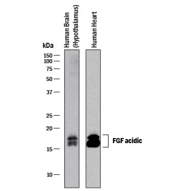
Detection of Human FGF acidic/FGF1 by Western Blot.
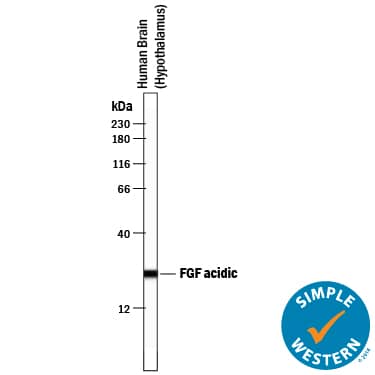
Western blot shows lysates of human brain (hypothalamas) tissue and human heart tissue. PVDF membrane was probed with 0.25 µg/mL of Goat Anti-Human FGF acidic/FGF1 Antigen Affinity-purified Polyclonal Antibody (Catalog # AF232) followed by HRP-conjugated Anti-Goat IgG Secondary Antibody (Catalog # HAF017). A specific band was detected for FGF acidic/FGF1 at approximately 16-17 kDa (as indicated). This experiment was conducted under reducing conditions and using Immunoblot Buffer Group 1.FGF acidic/FGF1 in Human Breast.
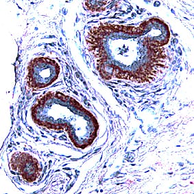
FGF acidic/FGF1 was detected in immersion fixed paraffin-embedded sections of human breast using Goat Anti-Human FGF acidic/FGF1 Antigen Affinity-purified Polyclonal Antibody (Catalog # AF232) at 15 µg/mL overnight at 4 °C. Tissue was stained using the Anti-Goat HRP-DAB Cell & Tissue Staining Kit (brown; Catalog # CTS008) and counterstained with hematoxylin (blue). Specific staining was localized to epithelial cells. View our protocol for Chromogenic IHC Staining of Paraffin-embedded Tissue Sections.Cell Proliferation Induced by FGF acidic/FGF1 and Neutralization by Human FGF acidic/FGF1 Antibody.
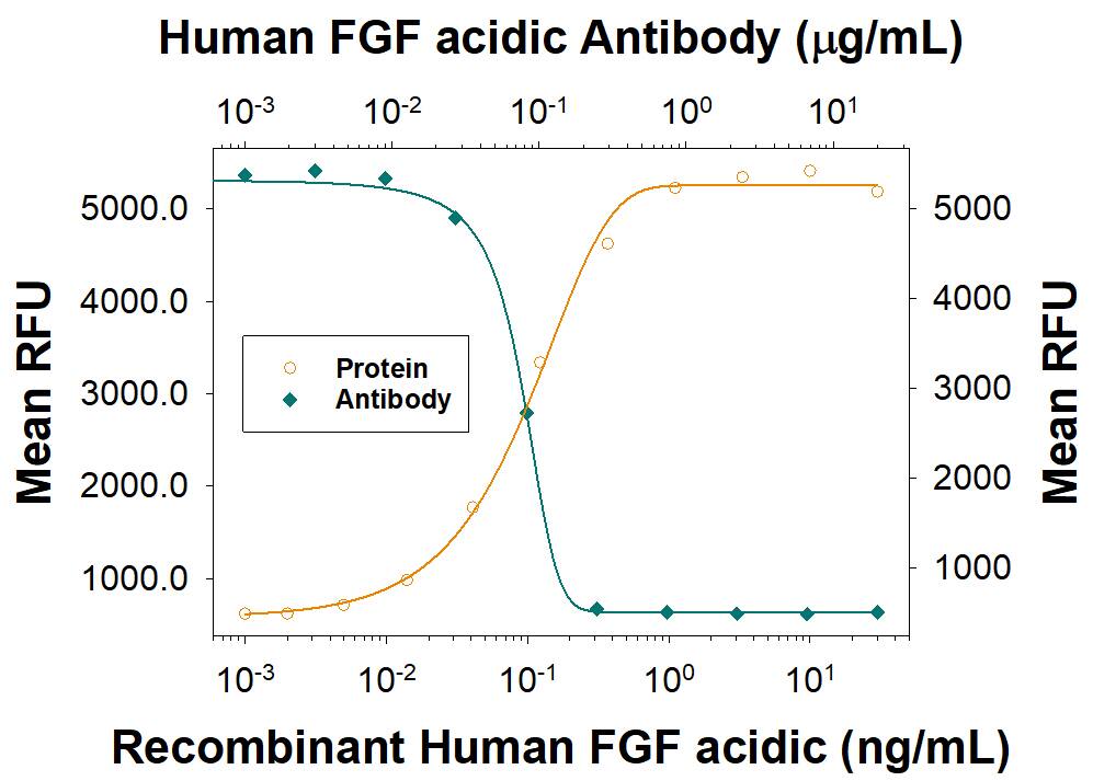
Recombinant Human FGF acidic/FGF1 aa 16-155 (Catalog # 232-FA) stimulates proliferation in the the NR6R-3T3 mouse fibroblast cell line in a dose-dependent manner (orange line). Proliferation elicited by Recombinant Human FGF acidic/FGF1 aa 16-155 (0.75 ng/mL) is neutralized (green line) by increasing concentrations of Goat Anti-Human FGF acidic/FGF1 Antigen Affinity-purified Polyclonal Antibody (Catalog # AF232). The ND50 is typically < 2 µg/mL in the presence of heparin (10 µg/mL).Applications for Human FGF acidic/FGF1 Antibody
Immunohistochemistry
Sample: Immersion fixed paraffin-embedded sections of human breast
Simple Western
Sample: Human brain (hypothalamus) tissue
Western Blot
Sample: Human brain (hypothalamas) tissue and human heart tissue
Neutralization
Formulation, Preparation, and Storage
Purification
Reconstitution
Formulation
Shipping
Stability & Storage
- 12 months from date of receipt, -20 to -70 °C as supplied.
- 1 month, 2 to 8 °C under sterile conditions after reconstitution.
- 6 months, -20 to -70 °C under sterile conditions after reconstitution.
Background: FGF acidic/FGF1
FGF acidic, also known as FGF1, ECGF, and HBGF-1, is a 17 kDa nonglycosylated member of the FGF family of mitogenic peptides. FGF acidic, which is produced by multiple cell types, stimulates the proliferation of all cells of mesodermal origin and many cells of neuroectodermal, ectodermal, and endodermal origin. It plays a number of roles in development, regeneration, and angiogenesis (1-3). Human FGF acidic shares 54% amino acid sequence identity with FGF basic and 17%‑33% with other human FGFs. It shares 92%, 96%, 96%, and 96% aa sequence identity with bovine, mouse, porcine, and rat FGF acidic, respectively, and exhibits considerable species crossreactivity. Alternate splicing generates a truncated isoform of human FGF acidic that consists of the N-terminal 40% of the molecule and functions as a receptor antagonist (4). During its nonclassical secretion, FGF acidic associates with S100A13, copper ions, and the C2A domain of synaptotagmin 1 (5). It is released extracellularly as a disulfide-linked homodimer and is stored in complex with extracellular heparan sulfate (6). The ability of heparan sulfate to bind FGF acidic is determined by its pattern of sulfation, and alterations in this pattern during embryogenesis thereby regulate FGF acidic bioactivity (7). The association of FGF acidic with heparan sulfate is a prerequisite for its subsequent interaction with FGF receptors (8, 9). Ligation triggers receptor dimerization, transphosphorylation, and internalization of receptor/FGF complexes (10). Internalized FGF acidic can translocate to the cytosol with the assistance of Hsp90 and then migrate to the nucleus by means of its two nuclear localization signals (11-13). The phosphorylation of FGF acidic by nuclear PKC delta triggers its active export to the cytosol where it is dephosphorylated and degraded (14, 15). Intracellular FGF acidic functions as a survival factor by inhibiting p53 activity and proapoptotic signaling (16).
References
- Jaye, M. et al. (1986) Science 233:541.
- Galzie, Z. et al. (1997) Biochem. Cell Biol. 75:669.
- Presta, M. et al. (2005) Cytokine Growth Factor Rev. 16:159.
- Yu, Y.L. et al. (1992) J. Exp. Med. 175:1073.
- Rajalingam, D. et al. (2007) Biochemistry 46:9225.
- Guerrini, M. et al. (2007) Curr. Pharm. Des. 13:2045.
- Allen, B.L. and A.C. Rapraeger (2003) J. Cell Biol. 163:637.
- Robinson, C.J. et al. (2005) J. Biol. Chem. 280:42274.
- Mohammadi, M. et al. (2005) Cytokine Growth Factor Rev. 16:107.
- Wiedlocha, A. and V. Sorensen (2004) Curr. Top. Microbiol. Immunol. 286:45.
- Wesche, J. et al. (2006) J. Biol. Chem. 281:11405.
- Imamura, T. et al. (1990) Science 249:1567.
- Wesche, J. et al. (2005) Biochemistry 44:6071.
- Wiedlocha, A. et al. (2005) Mol. Biol. Cell 16:794.
- Nilsen, T. et al. (2007) J. Biol. Chem. 282:26245.
- Bouleau, S. et al. (2005) Oncogene 24:7839.
Long Name
Alternate Names
Gene Symbol
UniProt
Additional FGF acidic/FGF1 Products
Product Documents for Human FGF acidic/FGF1 Antibody
Product Specific Notices for Human FGF acidic/FGF1 Antibody
For research use only
