Human FOLR2 Antibody
R&D Systems, part of Bio-Techne | Catalog # AF5697


Key Product Details
Species Reactivity
Validated:
Human
Cited:
Human
Applications
Validated:
Immunocytochemistry, Western Blot
Cited:
Western Blot
Label
Unconjugated
Antibody Source
Polyclonal Sheep IgG
Product Specifications
Immunogen
Chinese hamster ovary cell line CHO-derived recombinant human FOLR2
Gln22-His228
Accession # P14207
Gln22-His228
Accession # P14207
Specificity
Detects human FOLR2 in direct ELISAs and Western blots. In direct ELISAs, less than 5% cross-reactivity with recombinant human (rh) FOLR1 and rhFOLR3 is observed.
Clonality
Polyclonal
Host
Sheep
Isotype
IgG
Scientific Data Images for Human FOLR2 Antibody
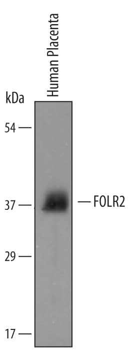
Detection of Human FOLR2 by Western Blot.
Western blot shows lysates of human placental tissue. PVDF membrane was probed with 1 µg/mL of Human FOLR2 Antigen Affinity-purified Polyclonal Antibody (Catalog # AF5697) followed by HRP-conjugated Anti-Sheep IgG Secondary Antibody (Catalog # HAF016). A specific band was detected for FOLR2 at approximately 38 kDa (as indicated). This experiment was conducted under reducing conditions and using Immunoblot Buffer Group 8.FOLR2 in Human Neutrophils.
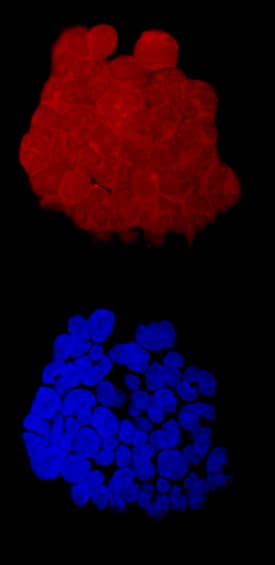
FOLR2 was detected in immersion fixed human neutrophils using Human FOLR2 Antigen Affinity-purified Polyclonal Antibody (Catalog # AF5697) at 10 µg/mL for 3 hours at room temperature. Cells were stained using the NorthernLights™ 557-conjugated Anti-Sheep IgG Secondary Antibody (red, upper panel; Catalog # NL010) and counterstained with DAPI (blue, lower panel). Specific staining was localized to cell surfaces and cytoplasm. View our protocol for Fluorescent ICC Staining of Non-adherent Cells.Applications for Human FOLR2 Antibody
Application
Recommended Usage
Immunocytochemistry
5-15 µg/mL
Sample: Immersion fixed human neutrophils
Sample: Immersion fixed human neutrophils
Western Blot
1 µg/mL
Sample: Human placental tissue
Sample: Human placental tissue
Reviewed Applications
Read 1 review rated 3 using AF5697 in the following applications:
Formulation, Preparation, and Storage
Purification
Antigen Affinity-purified
Reconstitution
Reconstitute at 0.2 mg/mL in sterile PBS. For liquid material, refer to CoA for concentration.
Formulation
Lyophilized from a 0.2 μm filtered solution in PBS with Trehalose. *Small pack size (SP) is supplied either lyophilized or as a 0.2 µm filtered solution in PBS.
Shipping
Lyophilized product is shipped at ambient temperature. Liquid small pack size (-SP) is shipped with polar packs. Upon receipt, store immediately at the temperature recommended below.
Stability & Storage
Use a manual defrost freezer and avoid repeated freeze-thaw cycles.
- 12 months from date of receipt, -20 to -70 °C as supplied.
- 1 month, 2 to 8 °C under sterile conditions after reconstitution.
- 6 months, -20 to -70 °C under sterile conditions after reconstitution.
Background: FOLR2
References
- Fowler, B. et al. (2001) Kidney Int. 59:S-221.
- Kelemen, L.E. (2006) Int. J. Cancer 119:243.
- Shen, F. et al. (1994) Biochemistry 33:1209.
- Ratnam, M. et al. (1989) Biochemistry 28:8249.
- Ross, J.F. et al. (1999) Cancer 85:348.
- Reddy, J.A. et al. (1999) Blood 93:3940.
- Ross, J.F. et al. (1994) Cancer 73:2432.
- Nakashima-Matsushita, N. et al. (1999) Arthritis Rheum. 42:1609.
- van der Heijden, J.W. et al. (2009) Arthritis Rheum. 60:12.
- Nagai, T. et al. (2009) Cancer Immunol. Immunother. Feb 24 epub.
- Piedrahita, J.A. et al. (1999) Nat. Genet. 23:228.
- Wlodarczyk, B. et al. (2001) Toxicol. Appl. Pharmacol. 177:238.
Long Name
Folate Receptor 2
Alternate Names
beta-HFR, FR-beta, FR-P3
Gene Symbol
FOLR2
UniProt
Additional FOLR2 Products
Product Documents for Human FOLR2 Antibody
Product Specific Notices for Human FOLR2 Antibody
For research use only
Loading...
Loading...
Loading...
Loading...
