Human Galectin-10 Antibody
R&D Systems, part of Bio-Techne | Catalog # MAB5447


Key Product Details
Species Reactivity
Applications
Label
Antibody Source
Product Specifications
Immunogen
Met1-Arg142
Accession # Q05315
Specificity
Clonality
Host
Isotype
Scientific Data Images for Human Galectin-10 Antibody
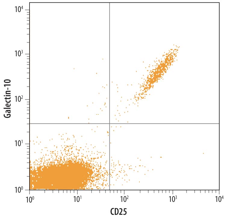
Detection of Galectin‑10 in Human PBMCs by Flow Cytometry.
Human peripheral blood lymphocytes were stained with Human Galectin-10 Monoclonal Antibody (Catalog # MAB5447) followed by Fluorescein-conjugated Anti-Mouse IgG Secondary Antibody (Catalog # F0103B) and Human IL-2 Ra APC-conjugated Monoclonal Antibody (Catalog # FAB1020A). Quadrant markers were set based on control antibody staining (Catalog # IC0041Fand IC003A).Detection of Human Galectin‑10 by Western Blot.
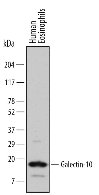
Western blot shows lysates of human eosinophils (enriched, approximately 60%). PVDF Membrane was probed with 1 µg/mL of Human Galectin-10 Monoclonal Antibody (Catalog # MAB5447) followed by HRP-conjugated Anti-Mouse IgG Secondary Antibody (Catalog # HAF007). A specific band was detected for Galectin-10 at approximately 16 kDa (as indicated). This experiment was conducted under reducing conditions and using Immunoblot Buffer Group 1.Galectin‑10 in HL‑60 Human Cell Line.
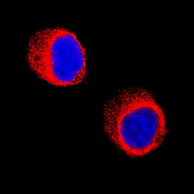
Galectin-10 was detected in immersion fixed HL-60 human acute promyelocytic leukemia cell line using Mouse Anti-Human Galectin-10 Monoclonal Antibody (Catalog # MAB5447) at 8 µg/mL for 3 hours at room temperature. Cells were stained using the NorthernLights™ 557-conjugated Anti-Mouse IgG Secondary Antibody (red; Catalog # NL007) and counterstained with DAPI (blue). Specific staining was localized to cytoplasm. View our protocol for Fluorescent ICC Staining of Non-adherent Cells.Applications for Human Galectin-10 Antibody
CyTOF-ready
Flow Cytometry
Sample: Human peripheral blood lymphocytes
Immunocytochemistry
Sample: Immersion fixed HL‑60 human acute promyelocytic leukemia cell line
Western Blot
Sample: Human eosinophils (enriched, approximately 60%)
Formulation, Preparation, and Storage
Purification
Reconstitution
Formulation
Shipping
Stability & Storage
- 12 months from date of receipt, -20 to -70 °C as supplied.
- 1 month, 2 to 8 °C under sterile conditions after reconstitution.
- 6 months, -20 to -70 °C under sterile conditions after reconstitution.
Background: Galectin-10
Galectin-10 (also eosinophil lysophospholipase and Charcot-Leyden Crystal protein) is a 16 kDa member of the lectin family of proteins. It is expressed intracellularly by eosinophils, basophils and CD25+ Treg cells. Although originally believed to possess lysophospholipase activity, this has been shown to be incorrect. It is known to bind lysophospholipase and its inhibitors, and to bind mannose in a very unusual manner. Human Galectin-10 is 142 amino acids (aa) in length. There is one galectin domain (aa 6-138) that contains two dimerization motifs (aa 6-10 and 131-135). Two molecular weight isoforms of 15 and 14 kDa have been described. Human Galectin-10 has no known structural counterpart in rodents.
Alternate Names
Entrez Gene IDs
Gene Symbol
UniProt
Additional Galectin-10 Products
Product Documents for Human Galectin-10 Antibody
Product Specific Notices for Human Galectin-10 Antibody
For research use only

