Human Galectin-3BP/MAC-2BP Antibody
R&D Systems, part of Bio-Techne | Catalog # AF2226

Key Product Details
Species Reactivity
Validated:
Cited:
Applications
Validated:
Cited:
Label
Antibody Source
Product Specifications
Immunogen
Val19-Asp585
Accession # Q08380
Specificity
Clonality
Host
Isotype
Scientific Data Images for Human Galectin-3BP/MAC-2BP Antibody
Detection of Human Galectin‑3BP/MAC‑2BP by Western Blot.
Western blot shows lysates of HeLa human cervical epithelial carcinoma cell line and HepG2 human hepatocellular carcinoma cell line. PVDF membrane was probed with 0.2 µg/mL of Goat Anti-Human Galectin-3BP/MAC-2BP Antigen Affinity-purified Polyclonal Antibody (Catalog # AF2226) followed by HRP-conjugated Anti-Goat IgG Secondary Antibody (Catalog # HAF019). A specific band was detected for Galectin-3BP/MAC-2BP at approximately 75 kDa (as indicated). This experiment was conducted under reducing conditions and using Immunoblot Buffer Group 1.Galectin‑3BP/MAC‑2BP in HeLa Human Cell Line.
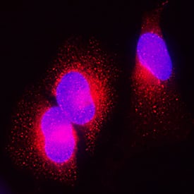
Galectin-3BP/MAC-2BP was detected in immersion fixed HeLa human cervical epithelial carcinoma cell line using Goat Anti-Human Galectin-3BP/MAC-2BP Antigen Affinity-purified Polyclonal Antibody (Catalog # AF2226) at 1.7 µg/mL for 3 hours at room temperature. Cells were stained using the NorthernLights™ 557-conjugated Anti-Goat IgG Secondary Antibody (red; Catalog # NL001) and counterstained with DAPI (blue). Specific staining was localized to cytoplasm. View our protocol for Fluorescent ICC Staining of Cells on Coverslips.Detection of Human Galectin‑3BP/MAC‑2BP by Simple WesternTM.
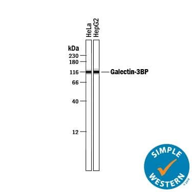
Simple Western lane view shows lysates of HeLa human cervical epithelial carcinoma cell line and HepG2 human hepatocellular carcinoma cell line, loaded at 0.2 mg/mL. A specific band was detected for Galectin-3BP/MAC-2BP at approximately 115 kDa (as indicated) using 10 µg/mL of Goat Anti-Human Galectin-3BP/MAC-2BP Antigen Affinity-purified Polyclonal Antibody (Catalog # AF2226) followed by 1:50 dilution of HRP-conjugated Anti-Goat IgG Secondary Antibody (Catalog # HAF109). This experiment was conducted under reducing conditions and using the 12-230 kDa separation system.Applications for Human Galectin-3BP/MAC-2BP Antibody
Immunocytochemistry
Sample: Immersion fixed HeLa human cervical epithelial carcinoma cell line
Simple Western
Sample: HeLa human cervical epithelial carcinoma cell line and HepG2 human hepatocellular carcinoma cell line
Western Blot
Sample: HeLa human cervical epithelial carcinoma cell line and HepG2 human hepatocellular carcinoma cell line
Reviewed Applications
Read 6 reviews rated 4 using AF2226 in the following applications:
Formulation, Preparation, and Storage
Purification
Reconstitution
Formulation
Shipping
Stability & Storage
- 12 months from date of receipt, -20 to -70 °C as supplied.
- 1 month, 2 to 8 °C under sterile conditions after reconstitution.
- 6 months, -20 to -70 °C under sterile conditions after reconstitution.
Background: Galectin-3BP/MAC-2BP
Galectin-3 binding protein (Galectin-3BP), also known as MAC-2 binding protein (MAC-2BP or M2BP), and the 90 kDa tumor associated antigen (TAA90K or 90K), is a secreted glycoprotein of the scavenger receptor cysteine-rich (SRCR) superfamily (1, 2). Galectin-3BP binds Galectin-3 (formerly MAC-2) with high affinity, but also binds Galectins -1 and -7, several collagen types, fibronectin, beta1 integrins and nidogen (3, 6, 7). It is widely expressed in all extracellular fluids and in pericellular areas of cell-rich tissues (1-3). The 585 amino acid (aa) human Galectin-3BP contains an 18 aa signal sequence and four definitive domains (4-6). Domain 1 is a group A scavenger receptor domain (4), domain 2 is a BTB/POZ domain that strongly mediates dimerization (5), and domain 3 is an IVR domain, that is also found following the POZ domain in Drosophila kelch protein. Although little is known about domain 4, recombinant domains 3 and 4 reproduce the solid-phase adhesion profile of full-length Galectin-3BP (5, 6). Glycosylation at seven N-linked sites, generates a molecular size of 85-97 kDa (1, 2, 6). Galectin-3BP dimers form linear and ring-shaped oligomers, most commonly decamers and dodecamers (3, 5). In vitro, Galectin-3BP has been shown to stimulate natural killer cells and lymphokine-activated killer cell activity (2). High Galectin-3BP expression has been correlated with tumor aggressiveness in several, but not all, study systems (7). Mature human Galectin-3BP shares 69% aa identity with mouse cyclophilin C-associated protein (CyCAP), which does not appear to bind Galectin-3 (8). Human Galectin-3BP also shares 73%, 67% and 68% aa identity with relatively uncharacterized orthologs in dog, rat and cow, respectively. A human N-terminally truncated sequence that begins within the BTB/POZ domain (aa 196) has been reported.
References
- Koths, K. et al. (1993) J. Biol. Chem. 268:14245.
- Ullrich, A. et al. (1994) J. Biol. Chem. 269:18401.
- Sasaki, T. et al. (1998) EMBO J. 17:1606.
- Hohenester, E. et al. (1999) Nat. Struct. Biol. 6:228.
- Muller, S. A. et al. (1999) J. Mol. Biol. 291:801.
- Hellstern, S. et al. (2002) J. Biol. Chem. 277:15690.
- Grassadonia, A. et al. (2004) Glycoconj. J. 19:551.
- Jalkanen, K. et al. (2001) Eur. J. Immunol. 31:3075.
Long Name
Alternate Names
Entrez Gene IDs
Gene Symbol
UniProt
Additional Galectin-3BP/MAC-2BP Products
Product Documents for Human Galectin-3BP/MAC-2BP Antibody
Product Specific Notices for Human Galectin-3BP/MAC-2BP Antibody
For research use only


