Human Glycoprotein V/CD42d Antibody
R&D Systems, part of Bio-Techne | Catalog # MAB4249


Key Product Details
Species Reactivity
Applications
Label
Antibody Source
Product Specifications
Immunogen
Gln17-Gly523
Accession # P40197
Specificity
Clonality
Host
Isotype
Scientific Data Images for Human Glycoprotein V/CD42d Antibody
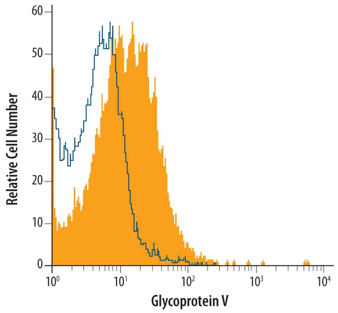
Detection of Glycoprotein V/CD42d in Human CD41+Platelets by Flow Cytometry.
Human blood-derived CD41+platelets were stained with Mouse Anti-Human Glycoprotein V/CD42d Mono-clonal Antibody (Catalog # MAB4249, filled histogram) or isotype control antibody (Catalog # MAB002, open histogram), followed by Allophycocyanin-conjugated Anti-Mouse IgG F(ab')2Secondary Antibody (Catalog # F0101B).Applications for Human Glycoprotein V/CD42d Antibody
CyTOF-ready
Flow Cytometry
Sample: Human blood-derived CD41+ platelets
Formulation, Preparation, and Storage
Purification
Reconstitution
Formulation
Shipping
Stability & Storage
- 12 months from date of receipt, -20 to -70 °C as supplied.
- 1 month, 2 to 8 °C under sterile conditions after reconstitution.
- 6 months, -20 to -70 °C under sterile conditions after reconstitution.
Background: Glycoprotein V/CD42d
GPV (platelet glycoprotein V; designated CD42d) is an 83 kDa type I transmembrane (TM) glycoprotein of the leucine‑rich repeat (LRR) family (1, 2). It is expressed exclusively within the platelet / megakaryocyte lineage, where it noncovalently interacts with other platelet TM LRR proteins, GPIb alpha/ beta and GPIX, at a ratio of one GPV to two of each other subunit (2). The GPI‑V‑IX complex tethers platelets to von Willebrand factor on the surface of injured endothelial cells. Absence of the complex results in Bernard‑Soulier syndrome, a rare bleeding disorder (1‑3). The human GPV cDNA encodes a 560 amino acid (aa) protein with a 16 aa signal sequence, a 507 aa extracellular domain (ECD) containing 15 LRR, a 21 aa TM sequence, and a short (16 aa) cytoplasmic tail that binds calmodulin in resting, but not activated platelets. The human GPV ECD shares 70%, 71% and 81% aa identity with mouse, rat and equine GPV, respectively. GPV can form soluble fragments of 80 kDa by ADAM10 or ADAM17 cleavage after P507, or 69 kDa by thrombin cleavage after R476 (1, 4, 5). High circulating soluble GPV may be an indicator of platelet activation, but may also be caused by high doses of aspirin (6‑8). The function of GPV is not entirely clear. Deletion of GPV in mice does not produce any obvious change to surface expression or function of GPIb and GPIX, but surface expression of GPV requires GPIb (9, 10). Deletion studies also indicate that GPV may play a minor role in collagen adhesion, and may modify platelet aggregation in response to thrombin (3, 11‑15).
References
- Lanza, F. et al. (1993) J. Biol. Chem. 268:20801.
- Hickey, M.J. et al. (1993) Proc. Natl. Acad. Sci. USA 90:8327.
- Ozaki, Y. et al. (2005) J. Thromb. Haemost. 3:1745.
- Rabie, T. et al. (2005) J. Biol. Chem. 280:14462.
- Gardiner, E.E. et al. (2007) J. Thromb. Haemost. 5:1530.
- Wolff, V. et al. (2005) Stroke 36:e17.
- Javela, K. et al. (2005) Transfusion 45:1504.
- Aktas, B. et al. (2005) J. Biol. Chem. 280:39716.
- Kahn, M.L. (1999) Blood 94:4112.
- Strassel, C. et al. (2004) Eur. J. Biochem. 271:3671.
- Nonne, C. et al. (2008) J. Thromb. Haemost. 6:210.
- Moog, S. et al. (2001) Blood 98:1038.
- Ramakrishnan, V. et al. (1999) Proc. Natl. Acad. Sci. USA 96:13336.
- Ni, H. et al. (2001) Blood 98:368.
- Andrews, R.K. et al. (2001) Blood 98:681.
Alternate Names
Gene Symbol
UniProt
Additional Glycoprotein V/CD42d Products
Product Documents for Human Glycoprotein V/CD42d Antibody
Product Specific Notices for Human Glycoprotein V/CD42d Antibody
For research use only