Human Glypican 6 Antibody
R&D Systems, part of Bio-Techne | Catalog # MAB2845


Key Product Details
Species Reactivity
Applications
Label
Antibody Source
Product Specifications
Immunogen
Asp24-Val527
Accession # Q9Y625
Specificity
Clonality
Host
Isotype
Scientific Data Images for Human Glypican 6 Antibody
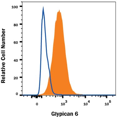
Detection of Glypican 6 in HepG2 Human Cell Line by Flow Cytometry.
HepG2 human hepatocellular carcinoma cell line was stained with Mouse Anti-Human Glypican 6 Monoclonal Antibody (Catalog # MAB2845, filled histogram) or isotype control antibody (Catalog # MAB002, open histogram), followed by Allophycocyanin-conjugated Anti-Mouse IgG Secondary Antibody (Catalog # F0101B). To facilitate intracellular staining, cells were fixed with Flow Cytometry Fixation Buffer (Catalog # FC004) and permeabilized with Flow Cytometry Permeabilization/Wash Buffer I (Catalog # FC005). View our protocol for Staining Intracellular Molecules.Applications for Human Glypican 6 Antibody
CyTOF-ready
Intracellular Staining by Flow Cytometry
Sample: HepG2 human hepatocellular carcinoma cell line fixed with Flow Cytometry Fixation Buffer (Catalog # FC004) and permeabilized with Flow Cytometry Permeabilization/Wash Buffer I (Catalog # FC005)
Western Blot
Sample: Recombinant Human Glypican 6 (Catalog # 2845-GP)
Formulation, Preparation, and Storage
Purification
Reconstitution
Formulation
Shipping
Stability & Storage
- 12 months from date of receipt, -20 to -70 °C as supplied.
- 1 month, 2 to 8 °C under sterile conditions after reconstitution.
- 6 months, -20 to -70 °C under sterile conditions after reconstitution.
Background: Glypican 6
The Glypicans (glypiated proteoglycans) are a small multigene family of GPI-linked heparan sulfate (HS) proteoglycans that likely play a key role in embryonic morphogenesis (1-4). There are currently six known mammalian Glypicans. They all share a common-sized protein core of 60‑70 kDa, an N-terminus which likely forms a compact globular domain, 14 conserved cysteines that form multiple intrachain disulfide bonds, and a number of C‑terminal N- and O-linked carbohydrate attachment sites. Based on exon organization and the location of O-linked glycosylation sites, at least two subfamilies of Glypicans are known, with one subfamily containing Glypicans 1, 2, 4 and 6, and another subfamily containing Glypicans 3 and 5 (3, 5). Human Glypican 6 (GPC-6) is synthesized as a 554 amino acid (aa) preproprecursor that contains a 23 aa signal sequence, a 505 aa mature region and a 26 aa C-terminal prosegment (5, 6). There are four consecutive Ser-Gly repeats that serve as a heparin sulfate attachment site. GPC-6 is reported to be as large as 110 kDa in size. This translates into approximately 50 kDa of proteoglycan (5). Human to mouse, there is 97% aa identity over the entire GPC-6 molecule. Cells known to express GPC-6 are adult ovary and embryonic vascular and visceral smooth muscle, plus mesenchyme (embryonic connective tissue) in multiple organs (1, 5, 6). The function of GPC-6 is essentially unknown. As a Glypican family member, it may facilitate heparin-binding growth factor signaling and polyamine uptake into expressing cells (7, 8). In this regard, it would appear that GPC-6 with its attendant HS is down‑regulated by triiodothyronine during cartilage maturation, thus limiting the availability of sites for FGF sequestration and activity (9).
References
- Song, H.H. and J. Filmus (2002) Biochim. Biophys. Acta 1573:241.
- Filmus, J. (2001) Glycobiology 11:19R.
- De Cat, B. and G. David (2001) Semin. Cell Dev. Biol. 12:117.
- Filmus, J. (2003) Glycoconj. J. 19:319.
- Veugelers, M. et al. (1999) J. Biol. Chem. 274:26968.
- Paine-Saunders, S. et al. (1999) Genomics 57:455.
- Fransson, L-A. et al. (2004) Cell Mol. Life Sci. 61:1016.
- Fransson, L-A. (2003) Int. J. Biochem. Cell Biol. 35:125.
- Bassett, J.H.D. et al. (2006) Endocrinology 147:295.
Alternate Names
Gene Symbol
UniProt
Additional Glypican 6 Products
Product Documents for Human Glypican 6 Antibody
Product Specific Notices for Human Glypican 6 Antibody
For research use only