Human IL-18 R alpha/IL-1 R5 Antibody
R&D Systems, part of Bio-Techne | Catalog # MAB840

Key Product Details
Validated by
Species Reactivity
Validated:
Cited:
Applications
Validated:
Cited:
Label
Antibody Source
Product Specifications
Immunogen
Glu20-Arg329
Accession # Q13478
Specificity
Clonality
Host
Isotype
Endotoxin Level
Scientific Data Images for Human IL-18 R alpha/IL-1 R5 Antibody
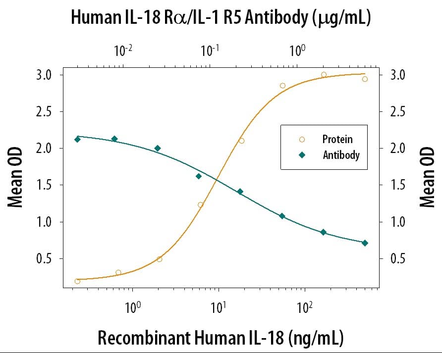
IFN‑ gamma Secretion Induced by IL‑18/IL‑1F4 and Neutralization by IL‑18 R alpha/IL‑1 R5 Antibody.
Human IL‑18 R alpha/IL‑1 R5 Antibody (Catalog # MAB840) neutralizes IL‑18/IL‑1F4 (9124-IL) induced IFN‑ gamma secretion in KG-1 human myeloid leukemia cells in the presence of Recombinant Human TNF-alpha (210-TA) as measured by the Human IFN-gamma Quantikine ELISA Kit (DIF50C). The Neutralization Dose (ND50) is typically < 0.500 µg/mL.IL-18 R alpha/IL-1 R5 in Human PBMCs.
IL-18 Ra/IL-1 R5 was detected in immersion fixed human peripheral blood mononuclear cells (PBMCs) using 10 µg/mL Mouse Anti-Human IL-18 Ra/IL-1 R5 Mono-clonal Antibody (Catalog # MAB840) for 3 hours at room temperature. Cells were stained with the NorthernLights™ 557-conjugated Anti-Mouse IgG Secondary Antibody (red; Catalog # NL007) and counter-stained with DAPI (blue). View our protocol for Fluorescent ICC Staining of Non-adherent Cells.Detection of IL-18 R alpha/IL-1 R5 in Human Skin.
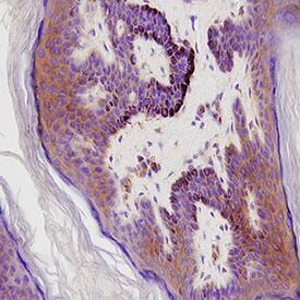
IL-18 R alpha/IL-1 R5 was detected in immersion fixed paraffin-embedded sections of Human Skin using Mouse Anti-Human IL-18 R alpha/IL-1 R5 Monoclonal Antibody (Catalog # MAB840) at 15 µg/mL for 1 hour at room temperature followed by incubation with the Anti-Mouse IgG VisUCyte™ HRP Polymer Antibody (Catalog # VC001). Before incubation with the primary antibody, tissue was subjected to heat-induced epitope retrieval using VisUCyte Antigen Retrieval Reagent-Basic (Catalog # VCTS021). Tissue was stained using DAB (brown) and counterstained with hematoxylin (blue). Specific staining was localized to cytoplasm and cell surface of keratinocytes. View our protocol for IHC Staining with VisUCyte HRP Polymer Detection Reagents.Applications for Human IL-18 R alpha/IL-1 R5 Antibody
CyTOF-ready
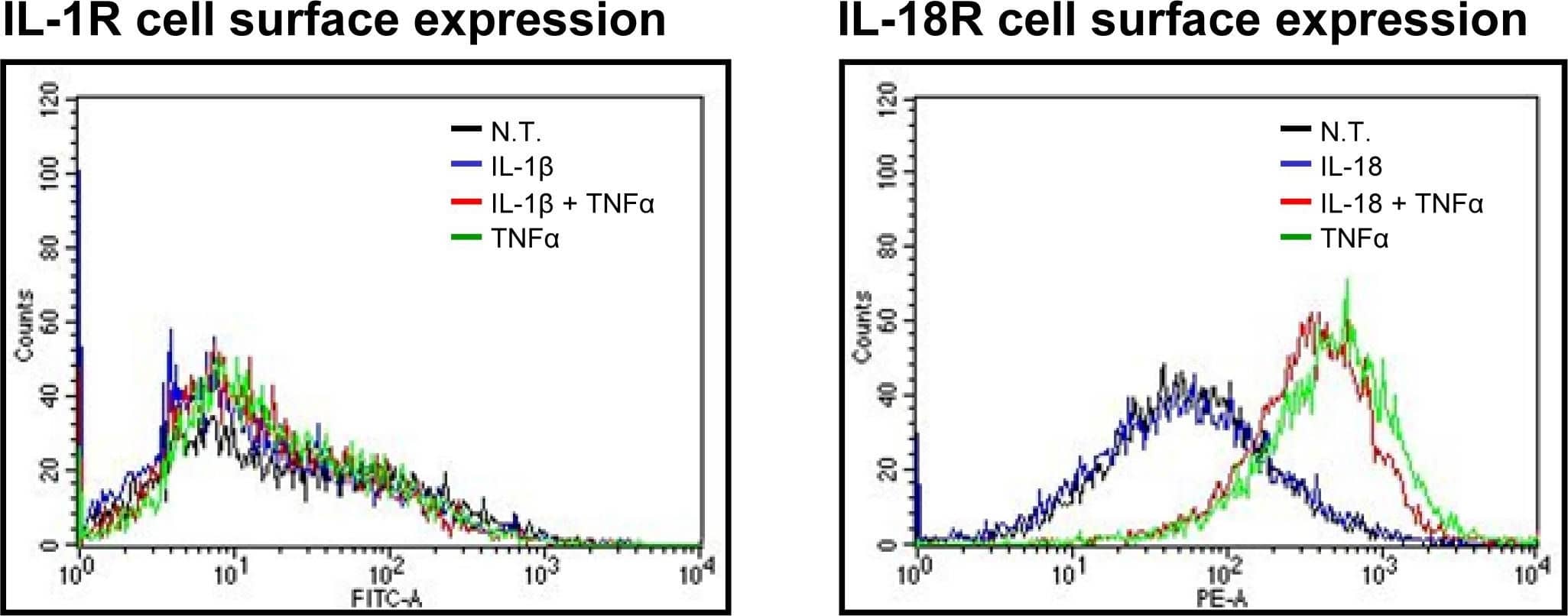
Flow Cytometry
Sample: Human peripheral blood mononuclear cells treated with PHA and Recombinant Human IL-2 (Catalog # 202-IL)
Immunocytochemistry
Sample: Immersion fixed human peripheral blood mononuclear cells (PBMCs)
Immunohistochemistry
Sample: Immersion fixed paraffin-embedded sections of human skin
Formulation, Preparation, and Storage
Purification
Reconstitution
Formulation
*Small pack size (-SP) is supplied either lyophilized or as a 0.2 µm filtered solution in PBS.
Shipping
Stability & Storage
- 12 months from date of receipt, -20 to -70 °C as supplied.
- 1 month, 2 to 8 °C under sterile conditions after reconstitution.
- 6 months, -20 to -70 °C under sterile conditions after reconstitution.
Background: IL-18 R alpha/IL-1 R5
Interleukin 18 (IL-18) is a member of the IL-1 family of cytokines and shares numerous immunoregulatory functions with IL-12. The functional IL-18 receptor complex is composed of two subunits designated IL-18 R alpha (also termed IL-1 R5 and IL-1 Rrp) and IL-18 R beta (also termed IL-1 R7 and AcPL). Both IL-18 R alpha and IL-18 R beta belong to the IL-1 receptor superfamily. Although IL-18 R by itself binds IL-18 with low affinity and IL-18 R beta does not bind IL-18 in vitro, co-expression of IL‑18 R alpha and IL‑18 R beta is required for high affinity binding and IL-18 responsiveness. Human IL-18 R cDNA encodes a 541 amino acid (aa) precursor type I membrane protein with a hydrophobic signal, an extracellular domain comprised of three immunoglobulin-like domains, a transmembrane domain and a cytoplasmic region of approximately 200 aa. Human and mouse IL-18 R share 65% amino acid sequence homology. IL-18 R is widely expressed in numerous tissues including spleen, thymus, leukocyte, liver, lung, heart, small and large intestine, prostate and placenta.
References
- Parnet, P. et al. (1996) J. Biol. Chem. 271:3967.
- Torigoe, K. et al. (1997) J. Biol. Chem. 272:25737.
- Born, T.L. et al. (1998) J. Biol. Chem. 273:29445.
Long Name
Alternate Names
Gene Symbol
UniProt
Additional IL-18 R alpha/IL-1 R5 Products
Product Documents for Human IL-18 R alpha/IL-1 R5 Antibody
Product Specific Notices for Human IL-18 R alpha/IL-1 R5 Antibody
For research use only

