Human IL-2 Antibody
R&D Systems, part of Bio-Techne | Catalog # AF-202-NA


Key Product Details
Validated by
Species Reactivity
Validated:
Cited:
Applications
Validated:
Cited:
Label
Antibody Source
Product Specifications
Immunogen
Ala21-Thr153
Accession # P60568
Specificity
Clonality
Host
Isotype
Endotoxin Level
Scientific Data Images for Human IL-2 Antibody
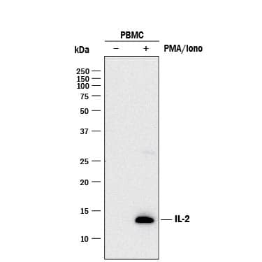
Detection of Human IL‑2 by Western Blot.
Western blot shows lysates of monensin treated human peripheral blood mononuclear cells (PBMCs) with no additional treatment (-) or additionally treated (+) with 0.5 μg/mL calcium ionomycin (Iono) and 50 ng/mL PMA overnight. PVDF membrane was probed with 0.5 µg/mL of Goat Anti-Human IL-2 Antigen Affinity-purified Polyclonal Antibody (Catalog # AF-202-NA) followed by HRP-conjugated Anti-Goat IgG Secondary Antibody (HAF017). A specific band was detected for IL-2 at approximately 14 kDa (as indicated). This experiment was conducted under reducing conditions and using Immunoblot Buffer Group 1.IL‑2 in Human PBMCs.
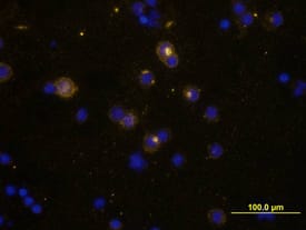
IL-2 was detected in immersion fixed human peripheral blood mononuclear cells (PBMCs) stimulated with PMA, ionomyocin, and monensin using Goat Anti-Human IL-2 Antigen Affinity-purified Polyclonal Antibody (Catalog # AF-202-NA) at 10 µg/mL for 3 hours at room temperature. Cells were stained using the NorthernLights™ 557-conjugated Anti-Goat IgG Secondary Antibody (yellow; (NL001) and counter-stained with DAPI (blue). View our protocol for Fluorescent ICC Staining of Non-adherent Cells.Cell Proliferation Induced by IL‑2 and Neutralization by Human IL‑2 Antibody.
Recombinant Human IL-2 (202-IL) stimulates proliferation in the CTLL-2 mouse cytotoxic T cell line in a dose-dependent manner (orange line) as measured by Resazurin (AR002). Proliferation elicited by Recombinant Human IL-2 (2 ng/mL) is neutralized (green line) by increasing concentrations of Goat Anti-Human IL-2 Antigen Affinity-purified Polyclonal Antibody (Catalog # AF-202-NA). The ND50 is typically ≤ 0.15 µg/mL.Applications for Human IL-2 Antibody
ELISA
This antibody functions as an ELISA detection antibody when paired with Mouse Anti-Human IL‑2 Monoclonal Antibody (Catalog # MAB2021).
This product is intended for assay development on various assay platforms requiring antibody pairs. We recommend the Human IL-2 DuoSet ELISA Kit (Catalog # DY202) for convenient development of a sandwich ELISA or the Human IL-2 Quantikine ELISA Kit (Catalog # D2050) for a complete optimized ELISA.
Immunocytochemistry
Sample: Immersion fixed human peripheral blood mononuclear cells stimulated with PMA, ionomycin, and monensin
Western Blot
Sample:
Human peripheral blood mononuclear cells (PBMCs) treated with monensin, 0.5ug/mL calcium ionomycin and 50ng/mL PMA overnight
Neutralization
Reviewed Applications
Read 2 reviews rated 5 using AF-202-NA in the following applications:
Formulation, Preparation, and Storage
Purification
Reconstitution
Formulation
Shipping
Stability & Storage
- 12 months from date of receipt, -20 to -70 °C as supplied.
- 1 month, 2 to 8 °C under sterile conditions after reconstitution.
- 6 months, -20 to -70 °C under sterile conditions after reconstitution.
Background: IL-2
Interleukin-2 (IL-2) is a O-glycosylated, four alpha-helix bundle cytokine that has potent stimulatory activity for antigen-activated T cells. It is expressed by CD4+ and CD8+ T cells, gamma delta T cells, B cells, dendritic cells, and eosinophils (1 - 3). Mature human IL-2 shares 56% and 66% aa sequence identity with mouse and rat IL-2, respectively. Human and mouse IL-2 exhibit cross-species activity (4). The receptor for IL-2 consists of three subunits that are present on the cell surface in varying preformed complexes (5 - 7). The 55 kDa IL-2 R alpha is specific for IL-2 and binds with low affinity. The 75 kDa IL-2 R beta, which is also a component of the IL-15 receptor, binds IL-2 with intermediate affinity. The 64 kDa common gamma chain gammac/IL-2 R gamma, which is shared with the receptors for IL-4, -7, -9, -15, and -21, does not independently interact with IL-2. Upon ligand binding, signal transduction is performed by both IL-2 R beta and gammac. IL-2 is best known for its autocrine and paracrine activity on T cells. It drives resting T cells to proliferate and induces IL-2 and IL-2 R alpha synthesis (1, 2). It contributes to T cell homeostasis by promoting the Fas-induced death of naïve CD4+ T cells but not activated CD4+ memory lymphocytes (8). IL-2 plays a central role in the expansion and maintenance of regulatory T cells, although it inhibits the development of Th17 polarized cells (9 - 11). Thus, IL-2 may be a key cytokine in the natural suppression of autoimmunity (12, 13).
References
- Ma, A. et al. (2006) Annu. Rev. Immunol. 24:657.
- Gaffen, S.L. and K.D. Liu (2004) Cytokine 28:109.
- Taniguchi, T. et al. (1983) Nature 302:305.
- Mosmann, T.R. et al. (1987) J. Immunol. 138:1813.
- Liparoto, S.F. et al. (2002) Biochemistry 41:2543.
- Wang, X. et al. (2005) Science 310:1159.
- Bodnar, A. et al. (2008) Immunol. Lett. 116:117.
- Jaleco, S. et al. (2003) J. Immunol. 171:61.
- Malek, T.R. (2003) J. Leukoc. Biol. 74:961.
- Laurence, A. et al. (2007) Immunity 26:371.
- Kryczek, I. et al. (2007) J. Immunol. 178:6730.
- Afzali, B. et al. (2007) Clin. Exp. Immunol. 148:32.
- Fehervari, Z. et al. (2006) Trends Immunol. 27:109.
Long Name
Alternate Names
Entrez Gene IDs
Gene Symbol
UniProt
Additional IL-2 Products
Product Documents for Human IL-2 Antibody
Product Specific Notices for Human IL-2 Antibody
For research use only

