Human IL-20R alpha Antibody
R&D Systems, part of Bio-Techne | Catalog # MAB11762


Key Product Details
Species Reactivity
Validated:
Cited:
Applications
Validated:
Cited:
Label
Antibody Source
Product Specifications
Immunogen
Val30-Lys250
Accession # Q9UHF4
Specificity
Clonality
Host
Isotype
Scientific Data Images for Human IL-20R alpha Antibody
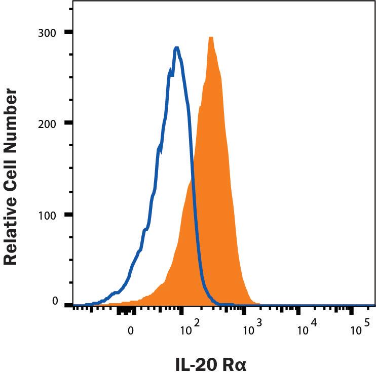
Detection of IL‑20 R alpha in T47D Cell Line by Flow Cytometry.
T47D human breast duct carcinoma cell line was stained with Mouse Anti-Human IL-20 Ra Monoclonal Antibody (Catalog # MAB11762, filled histogram) or isotype control antibody (MAB002, open histogram) followed by Anti-Mouse IgG PE-conjugated Secondary Antibody (F0102B). Staining was performed using our View our Staining Membrane-associated Proteins protocol.Applications for Human IL-20R alpha Antibody
CyTOF-ready
Flow Cytometry
Sample: Human IL‑20 R alpha transfected Baf/3 cells and T47D human breast carcinoma cell line
Western Blot
Sample: Recombinant Human IL-20 R alpha Fc Chimera (Catalog # 1176-IR)
Formulation, Preparation, and Storage
Purification
Reconstitution
Formulation
Shipping
Stability & Storage
- 12 months from date of receipt, -20 to -70 °C as supplied.
- 1 month, 2 to 8 °C under sterile conditions after reconstitution.
- 6 months, -20 to -70 °C under sterile conditions after reconstitution.
Background: IL-20R alpha
IL-20 receptor alpha (IL-20 R alpha), also named IL-20 R1, CRF2-8, and ZCYTOR7, belongs to the class II cytokine receptor family, which includes 12 members. These receptors are characterized by the patterns of conserved amino acid (aa) residues in their extracellular domains, which are composed of tandem fibronectin type III domains (1). Class II cytokine receptors form heterodimeric signaling receptor complexes that mediate class II cytokine signals. Subunits of the different receptor complexes are shared and serve multiple functions (1).
The gene for human IL-20 R alpha is mapped to chromosome 6 and encodes a 553 aa glycoprotein with a 29 aa signal peptide, a 221 aa extracellular domain, a 24 aa transmembrane region and a 279 aa intracellular domain (2). IL-20 R alpha is widely expressed and is detected at high levels in multiple tissues including skin, testis, heart, placenta, salivary gland and prostate gland (1). The expression of IL-20 R alpha, together with that of IL-20 R beta, is upregulated in psoriatic skin lesions on keratinocytes, immune cells, and endothelial cells (1, 2).
IL-20 R alpha heterodimerizes with IL-20 R beta to form the functional receptor that mediates IL-19, IL-20 and IL-24 signals (3, 4). IL-20 R alpha also heterodimerizes with IL-10 R beta to form the functional receptor complex for IL-26 (5). Binding of these IL-10 family class II cytokines to their functional receptors induces activation of the JAK-STAT signal transduction pathway. At low ligand concentrations, STAT3 has been shown to be the predominant STAT proteins activated through either complexes (3‑5).
References
- Kotenko, S.V. (2003) Cytokine & Growth Factor Reviews 13:223.
- Xie, M.H. et al. (2000) J. Biol. Chem. 275:31335.
- Dumoutier, L. et al. (2001) J. Immunol. 167:3534.
- Parrish-Novak, J. et al. (2002) J. Biol. Chem. 277:47517s.
- Sheikh. F. et al. (2004) J. Immunol. 172:2006.
Long Name
Alternate Names
Gene Symbol
UniProt
Additional IL-20R alpha Products
Product Documents for Human IL-20R alpha Antibody
Product Specific Notices for Human IL-20R alpha Antibody
For research use only