Human L1CAM Antibody
R&D Systems, part of Bio-Techne | Catalog # MAB7771


Key Product Details
Species Reactivity
Applications
Label
Antibody Source
Product Specifications
Immunogen
Accession # P32004
Specificity
Clonality
Host
Isotype
Scientific Data Images for Human L1CAM Antibody
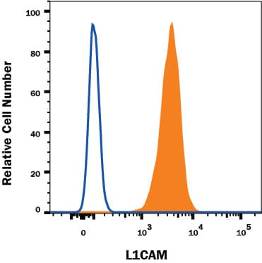
Detection of L1CAM in HeLa Human Cell Line by Flow Cytometry.
HeLa human cervical carcinoma cell line was stained with Mouse Anti-Human L1CAM Monoclonal Antibody (Catalog # MAB7771, filled histogram) or isotype control antibody (Catalog # MAB002, open histogram), followed by PE-conjugated Anti-Mouse IgG Secondary Antibody (Catalog # F0102B). View our protocol for Staining Membrane-associated Proteins.L1CAM in Human Brain (Cerebellum).
L1CAM was detected in immersion fixed paraffin-embedded sections of human brain (cerebellum) using Mouse Anti-Human L1CAM Monoclonal Antibody (Catalog # MAB7771) at 5 µg/mL for 1 hour at room temperature followed by incubation with the Anti-Mouse IgG VisUCyte™ HRP Polymer Antibody (Catalog # VC001). Before incubation with the primary antibody, tissue was subjected to heat-induced epitope retrieval using Antigen Retrieval Reagent-Basic (Catalog # CTS013). Tissue was stained using DAB (brown) and counterstained with hematoxylin (blue). Specific staining was localized to neuronal processes. View our protocol for IHC Staining with VisUCyte HRP Polymer Detection Reagents.L1CAM in Human Kidney Tissue.
L1CAM was detected in immersion fixed paraffin-embedded sections of human kidney tissue using Mouse Anti-Human L1CAM Monoclonal Antibody (Catalog # MAB7771) at 5 µg/mL for 1 hour at room temperature followed by incubation with the Anti-Mouse IgG VisUCyte™ HRP Polymer Antibody (Catalog # VC001). Before incubation with the primary antibody, tissue was subjected to heat-induced epitope retrieval using Antigen Retrieval Reagent-Basic (Catalog # CTS013). Tissue was stained using DAB (brown) and counterstained with hematoxylin (blue). Specific staining was localized to cell membrane in epithelial cells in convoluted tubules. View our protocol for IHC Staining with VisUCyte HRP Polymer Detection Reagents.Applications for Human L1CAM Antibody
CyTOF-ready
Flow Cytometry
Sample: HeLa human cervical carcinoma cell line
Immunohistochemistry
Sample: Immersion fixed paraffin-embedded sections of human brain (cerebellum) tissue and human kidney tissue
Formulation, Preparation, and Storage
Purification
Reconstitution
Formulation
Shipping
Stability & Storage
- 12 months from date of receipt, -20 to -70 °C as supplied.
- 1 month, 2 to 8 °C under sterile conditions after reconstitution.
- 6 months, -20 to -70 °C under sterile conditions after reconstitution.
Background: L1CAM
L1CAM (Neural cell adhesion molecule L1, also known as L1, CD171 and NCAM-L1) is a 200-230 kDa member of the L1 family, Immunoglobulin (Ig) superfamily of molecules. L1 is recognized to play a key role in cell migration, adhesion, neurite outgrowth, myelination and neuronal differentiation. It does so through a series of cis and trans interactions that involve multiple copartners and target receptors. L1 is described as forming both homotypic and heterotypic complexes, the latter with molecules as diverse as the EGFR, NCAM, CD24, neurocan and various alpha v plus beta 1 and beta 3 integrins. Cells known to express L1 include immature oligodendrocytes, CD4+ T cells, B cells and monocytes, pre-myelinating Schwann cells, intestinal epithelial progenitor cells, and cerebellar granule plus Purkinje cells. Mature human L1 is a 1238 amino acid (aa) type I transmembrane protein. It contains an 1101 aa extracellular region (aa 20-1120) plus a 114 aa cytoplasmic domain (aa 1144-1257). In general, the full-length L1 molecule is a neuron-associated isoform. L1 is known to undergo proteolysis, either by plasmin or ADAMs. This generates soluble isoforms of varying sizes (140-200 kDa) that retain bioactivity, and which can be incorporated into the surrounding ECM. The membrane fragments (30-80 kDa) undergo further processing, most importantly by gamma-secretase, to generate a soluble 28 kDa intracellular domain. This domain is SUMOylated, and believed to possess an NLS at Lys1147. Upon presumed entry into the nucleus, L1 is posited to activate L1-responsive genes. In the extracellular region, human and mouse L1 share 86% aa sequence identity.
Long Name
Alternate Names
Gene Symbol
UniProt
Additional L1CAM Products
Product Documents for Human L1CAM Antibody
Product Specific Notices for Human L1CAM Antibody
For research use only

