Human LAP (TGF-beta 1) Antibody
R&D Systems, part of Bio-Techne | Catalog # AF-246-NA


Key Product Details
Species Reactivity
Validated:
Cited:
Applications
Validated:
Cited:
Label
Antibody Source
Product Specifications
Immunogen
Leu30-Ser390
Accession # P01137
Specificity
Clonality
Host
Isotype
Endotoxin Level
Scientific Data Images for Human LAP (TGF-beta 1) Antibody
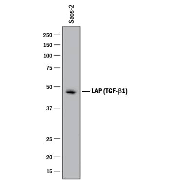
Detection of Human LAP (TGF-beta 1) by Western Blot.
Western blot shows lysates of Saos-2 human osteosarcoma cell line. PVDF membrane was probed with 2 µg/mL of Goat Anti-Human LAP (TGF-beta 1) Antigen Affinity-purified Polyclonal Antibody (Catalog # AF-246-NA) followed by HRP-conjugated Anti-Goat IgG Secondary Antibody (Catalog # HAF017). A specific band was detected for LAP (TGF-beta 1) at approximately 48 kDa (as indicated). This experiment was conducted under reducing conditions and using Immunoblot Buffer Group 1.LAP (TGF-beta 1) in Human PBMCs.
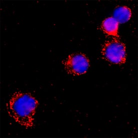
LAP (TGF-beta 1) was detected in immersion fixed Human peripheral blood mononuclear cells using Goat Anti-Human LAP (TGF-beta 1) Antigen Affinity-purified Polyclonal Antibody (Catalog # AF-246-NA) at 15 µg/mL for 3 hours at room temperature. Cells were stained using the NorthernLights™ 557-conjugated Anti-Goat IgG Secondary Antibody (red; NL001) and counterstained with DAPI (blue). Specific staining was localized to cytoplasm. Staining was performed using our protocol for Fluorescent ICC Staining of Non-adherent Cells.Detection of Human LAP (TGF-beta 1) by Simple WesternTM.
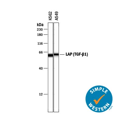
Simple Western lane view shows lysates of K562 human chronic myelogenous leukemia cell line and A549 human lung carcinoma cell line, loaded at 0.2 mg/mL. A specific band was detected for LAP (TGF-beta 1) at approximately 61 kDa (as indicated) using 20 µg/mL of Goat Anti-Human LAP (TGF-beta 1) Antigen Affinity-purified Polyclonal Antibody (Catalog # AF-246-NA) followed by 1:50 dilution of HRP-conjugated Anti-Goat IgG Secondary Antibody (Catalog # HAF109). This experiment was conducted under reducing conditions and using the 12-230 kDa separation system.Applications for Human LAP (TGF-beta 1) Antibody
Immunocytochemistry
Sample: Immersion fixed Human peripheral blood mononuclear cells
Simple Western
Sample: K562 human chronic myelogenous leukemia cell line and A549 human lung carcinoma cell line
Western Blot
Sample: Saos‑2 human osteosarcoma cell line
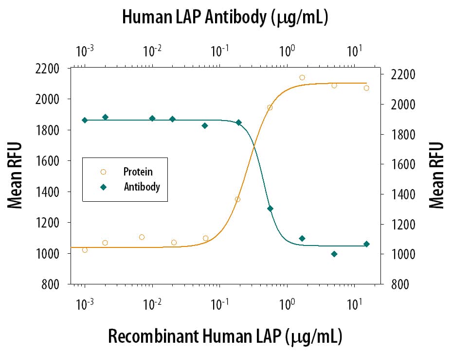
Neutralization
Reviewed Applications
Read 4 reviews rated 4 using AF-246-NA in the following applications:
Formulation, Preparation, and Storage
Purification
Reconstitution
Formulation
Shipping
Stability & Storage
- 12 months from date of receipt, -20 to -70 °C as supplied.
- 1 month, 2 to 8 °C under sterile conditions after reconstitution.
- 6 months, -20 to -70 °C under sterile conditions after reconstitution.
Background: LAP (TGF-beta 1)
TGF-beta 1 (transforming growth factor beta 1) and the closely related TGF-beta 2 and -beta 3 are members of the large TGF-beta superfamily. TGF‑ beta proteins are highly pleiotropic cytokines that regulate processes such as immune function, proliferation and epithelial-mesenchymal transition (1‑3). Human TGF-beta 1 cDNA encodes a 390 amino acid (aa) precursor that contains a 29 aa signal peptide and a 361 aa proprotein (4). A furin-like convertase processes the proprotein within the trans-Golgi to generate an N‑terminal 249 aa (aa 30-278) latency-associated peptide (LAP) and a C-terminal 112 aa mature TGF-beta 1 (aa 279-390) (4‑6). Disulfide-linked homodimers of LAP and TGF-beta 1 remain non‑covalently associated after secretion, forming the small latent TGF-beta 1 complex (4‑8). Purified LAP is also capable of associating with active TGF-beta with high affinity, and can neutralize TGF-beta activity (9). Covalent linkage of LAP to one of three latent TGF-beta binding proteins (LTBPs) creates a large latent complex that may interact with the extracellular matrix (5-7). TGF-beta activation from latency is controlled both spatially and temporally, by multiple pathways that include actions of proteases such as plasmin and MMP9, and/or by thrombospondin 1 or selected integrins (5, 8). The LAP portion of human TGF-beta 1 shares 91%, 92%, 85%, 86% and 88% aa identity with porcine, canine, mouse, rat and equine TGF-beta 1 LAP, respectively, while mature human TGF-beta 1 portion shares 100% aa identity with porcine, canine and bovine TGF-beta 1, and 99% aa identity with mouse, rat and equine TGF-beta 1. Although different isoforms of TGF-beta are naturally associated with their own distinct LAPs, the TGF-beta 1 LAP is capable of complexing with, and inactivating, all other human TGF-beta isoforms and those of most other species (9). Mutations within the LAP are associated with Camurati-Engelmann disease, a rare sclerosing bone dysplasia characterized by inappropriate presence of active TGF-beta 1 (10).
References
- Dunker, N. & K. Krieglstein (2000) Eur. J. Biochem. 267:6982.
- Wahl, S.M. (2006) Immunol. Rev. 213:213.
- Chang, H. et al. (2002) Endocr. Rev. 23:787.
- Derynck, R. et al. (1985) Nature 316:701.
- Dabovic, B. and D.B. Rifkin (2008) “TGF-beta Bioavailability” in The TGF-beta Family. Derynck, R. and K. Miyazono (eds): Cold Spring Harbor Laboratory Press, p. 179.
- Brunner, A.M. et al. (1989) J. Biol. Chem. 264:13660.
- Miyazono, K. et al. (1991) EMBO J. 10:1091.
- Oklu, R. and R. Hesketh (2000) Biochem. J. 352:601.
- Miller, D.M. et al. (1992) Mol. Endocrinol. 6:694.
- Janssens, K. et al. (2003) J. Biol. Chem. 278:7718.
Long Name
Alternate Names
Entrez Gene IDs
Gene Symbol
UniProt
Additional LAP (TGF-beta 1) Products
Product Documents for Human LAP (TGF-beta 1) Antibody
Product Specific Notices for Human LAP (TGF-beta 1) Antibody
For research use only


