Human LAP TGF-beta 1 Biotinylated Antibody
R&D Systems, part of Bio-Techne | Catalog # BAF246


Key Product Details
Species Reactivity
Validated:
Cited:
Applications
Validated:
Cited:
Label
Antibody Source
Product Specifications
Immunogen
Leu30-Ser390
Accession # P01137
Specificity
Clonality
Host
Isotype
Scientific Data Images for Human LAP TGF-beta 1 Biotinylated Antibody
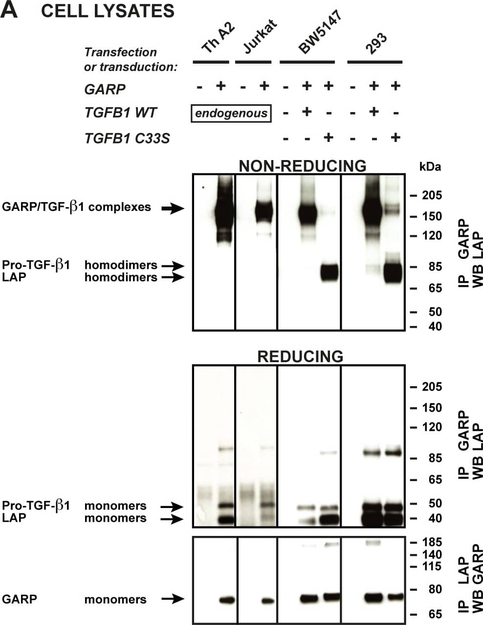
Detection of Human LAP (TGF-beta 1) by Western Blot
Disulfide-linked GARP/TGF-beta 1 complexes are released in the supernatant of T cells, but not 293 cells.A. Cells described in Figure 2 were lysed and immunoprecipitated (IP) with anti-GARP or anti-LAP antibodies. IP products were submitted to SDS-PAGE under non-reducing or reducing conditions, followed by WB with anti-LAP antibodies (top and middle panels), or anti-GARP antibodies (bottom panels). Pro-TGF-beta 1 and LAP homodimers in the top panels are not clearly resolved, but can be distinguished better with longer migrations or higher concentrations of polyacrylamide. The +/- 85-90 kDa bands that appear in the middle panel correspond to non-specific bands, or to incompletely reduced pro-TGF-beta 1. B. Cells (2x10⁶/ml for murine and human T cells, 2.5x10⁵/ml for transfected 293 cells) were incubated in serum free medium during 24 hours. Different cell concentrations were used to adjust for the different amounts of secreted TGF-beta 1 (see Figure 2). Human Th A2 and Jurkat cells were stimulated with anti-CD3/CD28 antibodies to increase secretion. Supernatants (0.5-10 µl) were analyzed by WB under non-reducing conditions with anti-GARP and anti-LAP antibodies. * Band that also appears when the secondary anti-IgG2b-HRP antibody is used alone (without anti-GARP antibody), due to cross reactivity against the anti-CD3/CD28 antibodies used for T cell stimulation. Image collected and cropped by CiteAb from the following publication (https://dx.plos.org/10.1371/journal.pone.0076186), licensed under a CC-BY license. Not internally tested by R&D Systems.Detection of Human LAP (TGF-beta 1) by Western Blot
Disulfide-linked GARP/TGF-beta 1 complexes are released by stimulated human Tregs, which naturally express GARP.The indicated Treg and Th cell populations were left resting or stimulated with anti-CD3/CD28 antibodies in serum-free medium. A. Supernatants were collected after 48 hours. B. Cell lysates were collected after 24 hours and IP with an anti-GARP antibody. Supernatants (A) and immunoprecipitated lysates (B) were submitted to SDS-PAGE under non-reducing conditions, then analyzed by WB with anti-LAP antibodies. Similar results were obtained with freshly isolated CD4+ T cells from 2 other donors and with expanded CD4+ T cells from 5 others donors. Image collected and cropped by CiteAb from the following publication (https://dx.plos.org/10.1371/journal.pone.0076186), licensed under a CC-BY license. Not internally tested by R&D Systems.Applications for Human LAP TGF-beta 1 Biotinylated Antibody
Western Blot
Sample: Recombinant Human LAP TGF-beta 1 (Catalog # 246-LP)
Formulation, Preparation, and Storage
Purification
Reconstitution
Formulation
Shipping
Stability & Storage
- 12 months from date of receipt, -20 to -70 °C as supplied.
- 1 month, 2 to 8 °C under sterile conditions after reconstitution.
- 6 months, -20 to -70 °C under sterile conditions after reconstitution.
Background: LAP (TGF-beta 1)
TGF-beta 1 (transforming growth factor beta 1) and the closely related TGF-beta 2 and -beta 3 are members of the large TGF-beta superfamily. TGF- beta proteins are highly pleiotropic cytokines that regulate processes such as immune function, proliferation and epithelial-mesenchymal transition (1-3). Human TGF-beta 1 cDNA encodes a 390 amino acid (aa) precursor that contains a 29 aa signal peptide and a 361 aa proprotein (4). A furin-like convertase processes the proprotein within the trans-Golgi to generate an N‑terminal 249 aa (aa 30-278) latency-associated peptide (LAP) and a C-terminal 112 aa (aa 279-390) mature TGF- beta1 (4-6). Disulfide-linked homodimers of LAP and TGF-beta 1 remain non‑covalently associated after secretion, forming the small latent TGF-beta 1 complex (4-8). Purified LAP is also capable of associating with active TGF-beta with high affinity, and can neutralize TGF-beta activity (9). Covalent linkage of LAP to one of three latent TGF-beta binding proteins (LTBPs) creates a large latent complex that may interact with the extracellular matrix (5‑7). TGF-beta activation from latency is controlled both spatially and temporally, by multiple pathways that include actions of proteases such as plasmin and MMP9, and/or by thrombospondin 1 or selected integrins (5, 8). The LAP portion of human TGF-beta 1 shares 91%, 92%, 85%, 86% and 88% aa identity with porcine, canine, mouse, rat and equine TGF-beta 1 LAP, respectively, while mature human TGF-beta 1 portion shares 100% aa identity with porcine, canine and bovine TGF-beta 1, and 99% aa identity with mouse, rat and equine TGF-beta 1. Although different isoforms of TGF-beta are naturally associated with their own distinct LAPs, the TGF-beta 1 LAP is capable of complexing with, and inactivating, all other human TGF-beta isoforms and those of most other species (9). Mutations within the LAP are associated with Camurati-Engelmann disease, a rare sclerosing bone dysplasia characterized by inappropriate presence of active TGF-beta 1 (10).
References
- Dunker, N. & K. Krieglstein (2000) Eur. J. Biochem. 267:6982.
- Wahl, S.M. (2006) Immunol. Rev. 213:213.
- Chang, H. et al. (2002) Endocr. Rev. 23:787.
- Derynck, R. et al. (1985) Nature 316:701.
- Dabovic, B. and D.B. Rifkin (2008) “TGF-beta Bioavailability” in The TGF-beta Family. Derynck, R. and K. Miyazono (eds): Cold Spring Harbor Laboratory Press, p. 179.
- Brunner, A.M. et al. (1989) J. Biol. Chem. 264:13660.
- Miyazono, K. et al. (1991) EMBO J. 10:1091.
- Oklu, R. and R. Hesketh (2000) Biochem. J. 352:601.
- Miller, D.M. et al. (1992) Mol. Endocrinol. 6:694.
- Janssens, K. et al. (2003) J. Biol. Chem. 278:7718.
Long Name
Alternate Names
Entrez Gene IDs
Gene Symbol
UniProt
Additional LAP (TGF-beta 1) Products
Product Documents for Human LAP TGF-beta 1 Biotinylated Antibody
Product Specific Notices for Human LAP TGF-beta 1 Biotinylated Antibody
For research use only
