Human LRIG3 Antibody
R&D Systems, part of Bio-Techne | Catalog # MAB3495


Key Product Details
Species Reactivity
Applications
Label
Antibody Source
Product Specifications
Immunogen
Asp28-Thr807
Accession # Q6UXM1
Specificity
Clonality
Host
Isotype
Scientific Data Images for Human LRIG3 Antibody
Detection of Human and Mouse LRIG3 by Western Blot.
Western blot shows lysates of KATO-III human gastric carcinoma cell line, HEK293T human embryonic kidney cell line, and B16-F1 mouse melanoma cell line. PVDF membrane was probed with 1 µg/mL of Mouse Anti-Human LRIG3 Monoclonal Antibody (Catalog # MAB3495) followed by HRP-conjugated Anti-Mouse IgG Secondary Antibody (Catalog # HAF007). A specific band was detected for LRIG3 at approximately 160 kDa (as indicated). This experiment was conducted under reducing conditions and using Immunoblot Buffer Group 1.Detection of LRIG3 in KATO‑III Human Cell Line by Flow Cytometry.
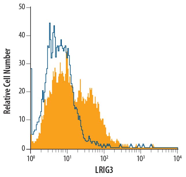
KATO-III human gastric carcinoma cell line was stained with Mouse Anti-Human LRIG3 Monoclonal Antibody (Catalog # MAB3495, filled histogram) or isotype control antibody (Catalog # MAB0041, open histogram), followed by Phycoerythrin-conjugated Anti-Mouse IgG Secondary Antibody (Catalog # F0102B).Applications for Human LRIG3 Antibody
CyTOF-ready
Flow Cytometry
Sample: KATO‑III human gastric carcinoma cell line
Western Blot
Sample: KATO‑III human gastric carcinoma cell line, HEK293T human embryonic kidney cell line, and B16‑F1 mouse melanoma cell line
Formulation, Preparation, and Storage
Purification
Reconstitution
Formulation
Shipping
Stability & Storage
- 12 months from date of receipt, -20 to -70 °C as supplied.
- 1 month, 2 to 8 °C under sterile conditions after reconstitution.
- 6 months, -20 to -70 °C under sterile conditions after reconstitution.
Background: LRIG3
LRIG3 (leucine-rich repeats and Ig-like domains-3) is an approximately 140-170 kDa type I transmembrane glycoprotein member of the mammalian LRIG glycoprotein family. This family contains three members who share 45 - 50% amino acid (aa) identity (1). All members contain at least fifteen LRRs, accompanied by two flanking cysteine-rich regions, and three C2-type Ig-like domains in their extracellular domains (ECD) (1). LRIG3 mRNA is widely expressed, with highest levels in stomach, skin, thyroid and small intestine (1). Human LRIG3 is synthesized as a 1120 amino acid (aa) precursor. It contains a 24 aa signal sequence, a 786 aa ECD, a 21 aa transmembrane sequence, and a 289 aa intracellular region. One splice variant exists that has a 19 aa substitution for the first 79 aa of the standard (or long) form. This substitution appears to encode an alternate signal sequence, resulting in a mature protein that lacks the first and part of the second LRR. LRIG1, a related family member, is known to bind the EGF family receptors ErbB1-4, via either its LRR or Ig-like domains. It also binds the ubiquitin ligase, c-Cbl, and promotes ubiquitination, internalization and destruction of these receptors (2, 3). It is not known whether LRIG3 performs similar functions. Within the cell, LRIG3 is expressed in the perinuclear region as well as on the cell surface. Perinuclear location of LRIG3 in grade III and IV astrocytic tumors has been associated with better patient survival (4). Human LRIG3 ECD shares 91%, 92%, 95% and 98% aa sequence identity with mouse, rat, bovine and canine LRIG3 ECD, respectively.
References
- Guo, D. et al. (2004) Genomics 84:157.
- Gur, G. et al. (2004) EMBO J. 23:3270.
- Laederich, M.B. (2004) J. Biol. Chem. 279:47050.
- Guo, D. et al. (2006) Acta Neuropathol. (Berl.) 111:238.
Long Name
Alternate Names
Gene Symbol
UniProt
Additional LRIG3 Products
Product Documents for Human LRIG3 Antibody
Product Specific Notices for Human LRIG3 Antibody
For research use only
