Human MICB Antibody
R&D Systems, part of Bio-Techne | Catalog # MAB1599


Key Product Details
Validated by
Species Reactivity
Validated:
Cited:
Applications
Validated:
Cited:
Label
Antibody Source
Product Specifications
Immunogen
Ala23-Gly298
Accession # CAI18747
Specificity
Clonality
Host
Isotype
Scientific Data Images for Human MICB Antibody
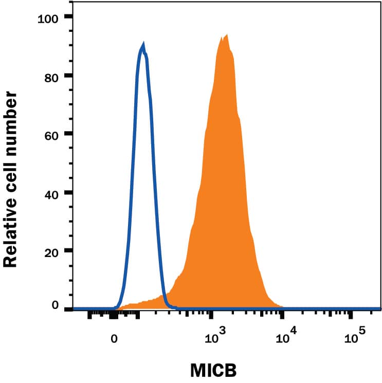
Detection of MICB in K562 Human Cell Line by Flow Cytometry.
K562 human chronic myelogenous leukemia cell line was stained with Mouse Anti-Human MICB Monoclonal Antibody (Catalog # MAB1599, filled histogram) or isotype control antibody (Catalog # MAB0041, open histogram), followed by Phycoerythrin-conjugated Anti-Mouse IgG Secondary Antibody (Catalog # F0102B).MICB Specificity is Shown by Flow Cytometry in Knockout Cell Line.
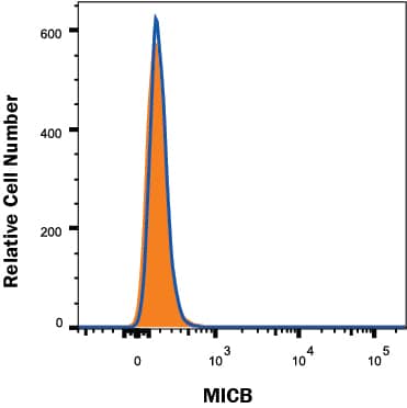
MICB knockout K562 human myelogenous leukemia cell line was stained with Mouse Anti-Human MICB Monoclonal Antibody (Catalog # MAB1599, filled histogram) or isotype control antibody (Catalog # MAB0041, open histogram) followed by anti-Mouse IgG PE-conjugated secondary antibody (Catalog # F0102B). No staining in the MICB knockout K562 cell line was observed. View our protocol for Staining Membrane-associated Proteins.Detection of Human MICB by Flow Cytometry
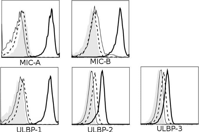
Multiple receptors and ligands are involved in NK cell-mediated lysis of activated CD4+ T cells.Role of (A) activating and (B) inhibitory NK receptors in NK cell degranulation. Left column: representative histograms (of n≥3) for surface expression of ligands on activated (thick black line) and resting CD4+ T cells (thin black line). Isotype-matched control Ig are represented by dashed line (activated CD4+ T) and filled histogram (resting CD4+ T). Middle- and right column: NK and CD4+ T cells were activated for 4 days in vitro as described, and co-cultured for 4 hours with 10 ug/mL mAb (or relevant isotype-matched control Ig). Degranulation is shown for CD56dim (middle column) and CD56bright (right column) NK cells. Representative histograms of surface expression of receptors on activated (thick black line) and resting NK cells (thin black line). Isotype-matched control Ig are represented by dashed line (activated NK) and filled histogram (resting NK). * P<0.05, ** P<0.005, *** P<0.001. (C) Sorted IL-2-activated CD56dim and CD56bright NK cells were co-cultured with 51Cr-labeled activated CD4+ T cells in a 51Cr-release assay with human IgG4 isotype control (•) or anti-NKG2A mAb (○). Data represents n = 3 experiments. Image collected and cropped by CiteAb from the following publication (https://pubmed.ncbi.nlm.nih.gov/22384114), licensed under a CC-BY license. Not internally tested by R&D Systems.Applications for Human MICB Antibody
CyTOF-ready
Flow Cytometry
Sample: K562 human chronic myelogenous leukemia cell line
Knockout Validated
Western Blot
Sample: Recombinant Human MICB Fc Chimera, aa 23-298 (Catalog # 1599-MB)
Human MICB Sandwich Immunoassay
Reviewed Applications
Read 1 review rated 5 using MAB1599 in the following applications:
Formulation, Preparation, and Storage
Purification
Reconstitution
Formulation
Shipping
Stability & Storage
- 12 months from date of receipt, -20 to -70 °C as supplied.
- 1 month, 2 to 8 °C under sterile conditions after reconstitution.
- 6 months, -20 to -70 °C under sterile conditions after reconstitution.
Background: MICB
MICB (MHC class I chain-related gene B) is a transmembrane glycoprotein that functions as a ligand for NKG2D. A closely related protein, MICA, shares 85% amino acid identity with MICB. These 2 proteins are distantly related to the MHC class I proteins. MICA and MICB (MICA/B) possess three extracellular immunoglobulin-like domains, but have no capacity to bind peptide or interact with beta2-microglobulin. The genes encoding MICA/B are found within the major histocompatibility complex on human chromosome 6. The MICB locus is polymorphic with more than 15 recognized human alleles. MICA/B are minimally expressed on normal cells, but are frequently expressed on epithelial tumors and can be induced by bacterial and viral infections. MICA/B are ligands for NKG2D, an activating receptor expressed on NK cells, NKT cells, gamma delta T cells, and CD8+ alpha beta T cells. Recognition of MICA/B by NKG2D results in the activation of cytolytic activity and/or cytokine production by these effector cells. MICA/B recognition is involved in tumor surveillance, viral infections, and autoimmune diseases. The release of soluble forms of MICA/B from tumors down-regulates NKG2D surface expression on effector cells resulting in the impairment of anti-tumor immune response (1-7).
References
- Groh, V. et al. (2001) Nature Immunol. 2:255.
- Stephens, H. (2001) Trends Immunol. 22:378.
- Bauer, S. et al. (1999) Science 285:727.
- Groh, V. et al. (2002) Nature 419:734.
- Steinle, A. et al. (2001) Immunogenetics 53:279.
- Pende, D. et al. (2002) Cancer Res. 62:6178.
- Salih, H. et al. (2003) Blood 102:1389.
Long Name
Alternate Names
Entrez Gene IDs
Gene Symbol
UniProt
Additional MICB Products
Product Documents for Human MICB Antibody
Product Specific Notices for Human MICB Antibody
For research use only


