Human MMP-10 Antibody
R&D Systems, part of Bio-Techne | Catalog # MAB9101

Key Product Details
Species Reactivity
Validated:
Cited:
Applications
Validated:
Cited:
Label
Antibody Source
Product Specifications
Immunogen
Tyr18-Cys476
Accession # P09238
Specificity
Clonality
Host
Isotype
Endotoxin Level
Scientific Data Images for Human MMP-10 Antibody
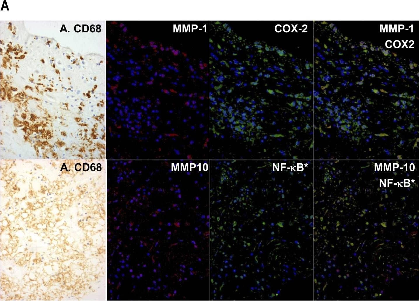
Detection of Human MMP-10 by Immunohistochemistry
Co-localisation of MMPs with markers of classical activation in human atherosclerotic plaques.(A) Serial sections were stained by peroxidase with anti-CD68 (A. CD68) and by dual immuno-fluorescence with anti-MMP-1 or anti-MMP-10 (red) together with anti-COX-2 or p65RelA (green) as shown, and the images were superimposed digitally. Nuclear localised p65RelA (NF-kB*) was detected because of the shift in nuclear counterstain colour from dark blue (DAPI alone) to sky blue (blue plus green) in the superimposed image. (B) Areas rich in macrophages identified from the peroxidase stain were identified in the serial section. Cells in the whole field were counted and a percentage of each staining pattern calculated. Values are means ± SEM, n = 6, * p<0.05. Image collected and cropped by CiteAb from the following open publication (https://pubmed.ncbi.nlm.nih.gov/22880008), licensed under a CC-BY license. Not internally tested by R&D Systems.Applications for Human MMP-10 Antibody
Immunohistochemistry
Sample: Immersion fixed paraffin-embedded sections of human intestine
Immunoprecipitation
Sample: Conditioned cell culture medium spiked with Recombinant Human MMP‑10 (Catalog # 910‑MP), see our available Western blot detection antibodies
Western Blot
Sample: Recombinant Human MMP-10 Western Blot Standard (Catalog # WBC026)
Reviewed Applications
Read 1 review rated 1 using MAB9101 in the following applications:
Formulation, Preparation, and Storage
Purification
Reconstitution
Formulation
Shipping
Stability & Storage
- 12 months from date of receipt, -20 to -70 °C as supplied.
- 1 month, 2 to 8 °C under sterile conditions after reconstitution.
- 6 months, -20 to -70 °C under sterile conditions after reconstitution.
Background: MMP-10
Matrix metalloproteinases are a family of zinc and calcium dependent endopeptidases with the combined ability to degrade all the components of the extracellular matrix. MMP-10 (stromelysin 2) degrades a broad range of substrates including gelatin, collagen types III, IV and V, fibronectin, aggrecan, and pig cartilage proteoglycan. MMP-10 can activate other MMPs such as MMP-1 and MMP-8. MMP-10 is expressed in keratinocytes, T cells, menstrual endometrium, and a few tumor samples. Structurally, MMP-10 may be divided into four distinct domains: a pro-domain which is cleaved upon activation, a catalytic domain containing the zinc binding site; a short linker region, and a carboxyl terminal hemopexin-like domain.
Long Name
Alternate Names
Entrez Gene IDs
Gene Symbol
UniProt
Additional MMP-10 Products
Product Documents for Human MMP-10 Antibody
Product Specific Notices for Human MMP-10 Antibody
For research use only
