Human/Mouse ATN1 Antibody
R&D Systems, part of Bio-Techne | Catalog # AF6567

Key Product Details
Species Reactivity
Applications
Label
Antibody Source
Product Specifications
Immunogen
Met1-Gln100
Accession # P54259
Specificity
Clonality
Host
Isotype
Scientific Data Images for Human/Mouse ATN1 Antibody
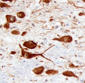
ATN1 in Human Brain.
ATN1 was detected in immersion fixed paraffin-embedded sections of human brain (hypothalamus) using Sheep Anti-Human/Mouse ATN1 Antigen Affinity-purified Polyclonal Antibody (Catalog # AF6567) at 3 µg/mL overnight at 4 °C. Before incubation with the primary antibody, tissue was subjected to heat-induced epitope retrieval using Antigen Retrieval Reagent-Basic (Catalog # CTS013). Tissue was stained using the Anti-Sheep HRP-DAB Cell & Tissue Staining Kit (brown; Catalog # CTS019) and counterstained with hematoxylin (blue). Specific staining was localized to neuronal cytoplasm. View our protocol for Chromogenic IHC Staining of Paraffin-embedded Tissue Sections.Detection of ATN1 in Mouse Liver.
ATN1 was detected in immersion fixed paraffin-embedded sections of mouse liver using Sheep Anti-Human/Mouse ATN1 Antigen Affinity-purified Polyclonal Antibody (Catalog # AF6567) at 1 µg/ml for 1 hour at room temperature followed by incubation with the Anti-Sheep IgG VisUCyte™ HRP Polymer Antibody (Catalog # VC006). Before incubation with the primary antibody, tissue was subjected to heat-induced epitope retrieval using VisUCyte Antigen Retrieval Reagent-Basic (Catalog # VCTS021). Tissue was stained using DAB (brown) and counterstained with hematoxylin (blue). Specific staining was localized to the nucleus. View our protocol for Chromogenic IHC Staining of Paraffin-embedded Tissue Sections.Applications for Human/Mouse ATN1 Antibody
Immunohistochemistry
Sample: Immersion fixed paraffin-embedded sections of human brain (hypothalamus)
Formulation, Preparation, and Storage
Purification
Reconstitution
Formulation
Shipping
Stability & Storage
- 12 months from date of receipt, -20 to -70 °C as supplied.
- 1 month, 2 to 8 °C under sterile conditions after reconstitution.
- 6 months, -20 to -70 °C under sterile conditions after reconstitution.
Background: ATN1
ATN1 (Atrophin-1; also DRPLA protein) is a 190‑200 kDa member of the Atrophin family of proteins that is cleaved into 120‑150 kDa fragments. ATN1 is a ubiquitously expressed transcriptional coactivator. Human ATN1 is 1185 amino acids (aa) in length. It contains multiple motifs, including an NLS (aa 16-32), interspersed poly-Pro, poly-Ser and poly-His regions (aa 376-707), two RE (ArgGlu) repeats (aa 816-934), and an NES (aa 1033-1042). There are at least 17 utilized phosphorylation sites and one acetylated Lys. ATN1 is most characterized by a poly-Gln region between aa 484-497. Normally, there are about 20 consecutive Gln residues, but this number may be increased to more than 70 in pathologic conditions. Proteolytic cleavage generates large C-terminal fragments of 120-150 kDa size. These are unlikely to contain the NLS, and thus are typically cytosolic. ATN1 is suggested to form heterodimers with full-length ATN2/RERE, thus generating a transcriptional repressor. There are multiple potential isoforms. One shows an alternative start site at Met527, while others differ in the number of glutamines in the poly-Gln region. Over aa 1-100, human ATN1 shares 94% aa identity with mouse ATN1.
Long Name
Alternate Names
Gene Symbol
UniProt
Additional ATN1 Products
Product Documents for Human/Mouse ATN1 Antibody
Product Specific Notices for Human/Mouse ATN1 Antibody
For research use only

