Human/Mouse Bax Antibody
R&D Systems, part of Bio-Techne | Catalog # AF820


Key Product Details
Validated by
Knockout/Knockdown
Species Reactivity
Validated:
Human, Mouse
Cited:
Human, Mouse, Rat
Applications
Validated:
Immunocytochemistry, Immunohistochemistry, Knockout Validated, Simple Western, Western Blot
Cited:
Immunohistochemistry-Paraffin, Western Blot
Label
Unconjugated
Antibody Source
Polyclonal Rabbit IgG
Product Specifications
Immunogen
KLH-coupled human Bax synthetic peptide
Specificity
Detects human and mouse Bax.
Clonality
Polyclonal
Host
Rabbit
Isotype
IgG
Scientific Data Images for Human/Mouse Bax Antibody
Detection of Human/Mouse Bax by Western Blot.
Western blot shows lysates of CHP-100 human neuroblastoma cell line, THP-1 human acute monocytic leukemia cell line, L-929 mouse fibroblast cell line, and DA3 mouse myeloma cell line. PVDF membrane was probed with 0.3 µg/mL of Rabbit Anti-Human/Mouse Bax Antigen Affinity-purified Polyclonal Antibody (Catalog # AF820) followed by HRP-conjugated Anti-Rabbit IgG Secondary Antibody (Catalog # HAF008). A specific band was detected for Bax at approximately 21 kDa (as indicated). This experiment was conducted under reducing conditions and using Immunoblot Buffer Group 2.Bax in A549 Human Cell Line.
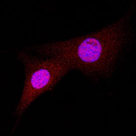
Bax was detected in immersion fixed A549 human lung carcinoma cell line using Rabbit Anti-Human/Mouse Bax Antigen Affinity-purified Polyclonal Antibody (Catalog # AF820) at 1.7 µg/mL for 3 hours at room temperature. Cells were stained using the NorthernLights™ 557-conjugated Anti-Rabbit IgG Secondary Antibody (red; Catalog # NL004) and counterstained with DAPI (blue). Specific staining was localized to cytoplasm and nuclei. View our protocol for Fluorescent ICC Staining of Cells on Coverslips.Bax in Human Breast.
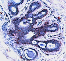
Bax was detected in immersion fixed paraffin-embedded sections of human breast using 15 µg/mL Rabbit Anti-Human/Mouse Bax Antigen Affinity-purified Polyclonal Antibody (Catalog # AF820) overnight at 4 °C. Tissue was stained (red) and counter-stained with hematoxylin (blue). View our protocol for Chromogenic IHC Staining of Paraffin-embedded Tissue Sections.Applications for Human/Mouse Bax Antibody
Application
Recommended Usage
Immunocytochemistry
1-15 µg/mL
Sample: Immersion fixed A549 human lung carcinoma cell line
Sample: Immersion fixed A549 human lung carcinoma cell line
Immunohistochemistry
5-15 µg/mL
Sample: Immersion fixed paraffin-embedded sections of human liver and immersion fixed paraffin-embedded sections of human breast
Sample: Immersion fixed paraffin-embedded sections of human liver and immersion fixed paraffin-embedded sections of human breast
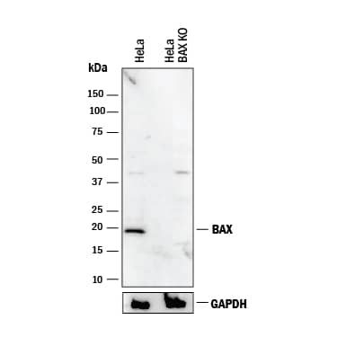
Knockout Validated
Bax is
specifically detected in HeLa human cervical epithelial carcinoma parental cell line but is not detectable in Bax
knockout HeLa cell line.
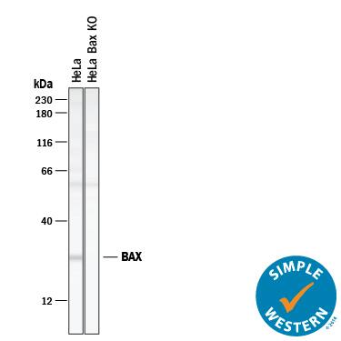
Simple Western
50 µg/mL
Sample: HeLa human cervical epithelial carcinoma cell line
Sample: HeLa human cervical epithelial carcinoma cell line
Western Blot
0.3 µg/mL
Sample: CHP-100 human neuroblastoma cell line, THP-1 human acute monocytic leukemia cell line, L-929 mouse fibroblast cell line, and DA3 mouse myeloma cell line
Sample: CHP-100 human neuroblastoma cell line, THP-1 human acute monocytic leukemia cell line, L-929 mouse fibroblast cell line, and DA3 mouse myeloma cell line
Reviewed Applications
Read 4 reviews rated 4.3 using AF820 in the following applications:
Formulation, Preparation, and Storage
Purification
Antigen Affinity-purified
Reconstitution
Reconstitute at 0.2 mg/mL in sterile PBS.
Formulation
Lyophilized from a 0.2 μm filtered solution in PBS with Trehalose.
Shipping
The product is shipped at ambient temperature. Upon receipt, store it immediately at the temperature recommended below.
Stability & Storage
Use a manual defrost freezer and avoid repeated freeze-thaw cycles.
- 12 months from date of receipt, -20 to -70 °C as supplied.
- 1 month, 2 to 8 °C under sterile conditions after reconstitution.
- 6 months, -20 to -70 °C under sterile conditions after reconstitution.
Background: Bax
Long Name
Bcl Associated X Protein
Alternate Names
apoptosis regulator BAX, BCL2-associated X protein, Bcl2-L-4, BCL2L4bcl2-L-4, Bcl-2-like protein 4
Gene Symbol
BAX
Additional Bax Products
Product Documents for Human/Mouse Bax Antibody
Product Specific Notices for Human/Mouse Bax Antibody
For research use only
Loading...
Loading...
Loading...
Loading...


