Human/Mouse Cadherin-13 Antibody
R&D Systems, part of Bio-Techne | Catalog # MAB3264


Conjugate
Catalog #
Key Product Details
Species Reactivity
Validated:
Human, Mouse
Cited:
Human, Mouse
Applications
Validated:
CyTOF-ready, Flow Cytometry, Immunohistochemistry, Western Blot
Cited:
Immunohistochemistry, Western Blot
Label
Unconjugated
Antibody Source
Monoclonal Rat IgG2A Clone # 392411
Product Specifications
Immunogen
Mouse myeloma cell line NS0-derived recombinant human Cadherin-13
Glu23-Ala692
Accession # P55290
Glu23-Ala692
Accession # P55290
Specificity
Detects human Cadherin-13 in direct ELISAs and Western blots. In direct ELISAs and Western blots, no cross-reactivity with recombinant human (rh) E-Cadherin, rhN-Cadherin, rhP-Cadherin, rhVE-Cadherin, rhCadherin-8, -11, -12 or -17 is observed.
Clonality
Monoclonal
Host
Rat
Isotype
IgG2A
Scientific Data Images for Human/Mouse Cadherin-13 Antibody
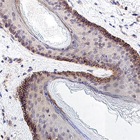
Detection of Cadherin-13 in Human Skin.
Cadherin-13 was detected in immersion fixed paraffin-embedded sections of human skin using Rat Anti-Human/Mouse Cadherin-13 Monoclonal Antibody (Catalog # MAB3264) at 5 µg/ml for 1 hour at room temperature followed by incubation with the HRP-conjugated Anti-Rat IgG Secondary Antibody (Catalog # HAF005) or the Anti-Rat IgG VisUCyte™ HRP Polymer Antibody (Catalog # VC005). Before incubation with the primary antibody, tissue was subjected to heat-induced epitope retrieval using VisUCyte Antigen Retrieval Reagent-Basic (Catalog # VCTS021). Tissue was stained using DAB (brown) and counterstained with hematoxylin (blue). Specific staining was localized to the membrane. View our protocol for Chromogenic IHC Staining of Paraffin-embedded Tissue Sections.Applications for Human/Mouse Cadherin-13 Antibody
Application
Recommended Usage
CyTOF-ready
Ready to be labeled using established conjugation methods. No BSA or other carrier proteins that could interfere with conjugation.
Flow Cytometry
0.25 µg/106 cells
Sample: NCI-H460 human large cell lung carcinoma cell line
Sample: NCI-H460 human large cell lung carcinoma cell line
Immunohistochemistry
3-25 µg/mL
Sample: Immersion fixed paraffin-embedded sections of human skin
Sample: Immersion fixed paraffin-embedded sections of human skin
Western Blot
1 µg/mL
Sample: Recombinant Human Cadherin‑13 under non-reducing conditions only (Catalog # 3264-CA)
Sample: Recombinant Human Cadherin‑13 under non-reducing conditions only (Catalog # 3264-CA)
Formulation, Preparation, and Storage
Purification
Protein A or G purified from hybridoma culture supernatant
Reconstitution
Reconstitute at 0.5 mg/mL in sterile PBS. For liquid material, refer to CoA for concentration.
Formulation
Lyophilized from a 0.2 μm filtered solution in PBS with Trehalose. See Certificate of Analysis for details.
*Small pack size (-SP) is supplied either lyophilized or as a 0.2 µm filtered solution in PBS.
*Small pack size (-SP) is supplied either lyophilized or as a 0.2 µm filtered solution in PBS.
Shipping
Lyophilized product is shipped at ambient temperature. Liquid small pack size (-SP) is shipped with polar packs. Upon receipt, store immediately at the temperature recommended below.
Stability & Storage
Use a manual defrost freezer and avoid repeated freeze-thaw cycles.
- 12 months from date of receipt, -20 to -70 °C as supplied.
- 1 month, 2 to 8 °C under sterile conditions after reconstitution.
- 6 months, -20 to -70 °C under sterile conditions after reconstitution.
Background: Cadherin-13
References
- Tanihara, H. et al. (1994) Cell Adhes. Commun. 2:15.
- Philippova, M. et al. (2009) Cell. Signal. 21:1035.
Alternate Names
Cadherin13, CDH13, H-Cadherin, T-Cadherin
Gene Symbol
CDH13
UniProt
Additional Cadherin-13 Products
Product Documents for Human/Mouse Cadherin-13 Antibody
Product Specific Notices for Human/Mouse Cadherin-13 Antibody
For research use only
Loading...
Loading...
Loading...
Loading...
Loading...