Human/Mouse HO-1/HMOX1/HSP32 Antibody
R&D Systems, part of Bio-Techne | Catalog # MAB3776

Key Product Details
Validated by
Species Reactivity
Validated:
Cited:
Applications
Validated:
Cited:
Label
Antibody Source
Product Specifications
Immunogen
Met1-Thr261
Accession # P09601
Specificity
Clonality
Host
Isotype
Scientific Data Images for Human/Mouse HO-1/HMOX1/HSP32 Antibody
Detection of Human/Mouse HO-1/HMOX1/HSP32 by Western Blot.
Western blot shows lysates of A549 human lung carcinoma cell line and A20 mouse B cell lymphoma cell line. PVDF membrane was probed with 1 µg/mL of Human/Mouse HO-1/HMOX1/HSP32 Monoclonal Antibody (Catalog # MAB3776) followed by HRP-conjugated Anti-Rat IgG Secondary Antibody (Catalog # HAF005). A specific band was detected for HO-1/HMOX1/HSP32 at approximately 32 kDa (as indicated). This experiment was conducted under reducing conditions and using Immunoblot Buffer Group 2.Western Blot Shows Human HO‑1/HMOX1/HSP32 Specificity by Using Knockout Cell Line.
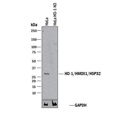
Western blot shows lysates of HeLa human cervical epithelial carcinoma parental cell line and HO-1/HMOX1/HSP32 knockout HeLa cell line (KO). PVDF membrane was probed with 1 µg/mL of Rat Anti-Human/Mouse HO-1/HMOX1/HSP32 Monoclonal Antibody (Catalog # MAB3776) followed by HRP-conjugated Anti-Rat IgG Secondary Antibody (Catalog # HAF005). A specific band was detected for HO-1/HMOX1/HSP32 at approximately 32 kDa (as indicated) in the parental HeLa cell line, but is not detectable in knockout HeLa cell line. GAPDH (Catalog # MAB5718) is shown as a loading control. This experiment was conducted under reducing conditions and using Immunoblot Buffer Group 1.Applications for Human/Mouse HO-1/HMOX1/HSP32 Antibody
Knockout Validated
Western Blot
Sample: A549 human lung carcinoma cell line and A20 mouse B cell lymphoma cell line
Formulation, Preparation, and Storage
Purification
Reconstitution
Formulation
Shipping
Stability & Storage
- 12 months from date of receipt, -20 to -70 °C as supplied.
- 1 month, 2 to 8 °C under sterile conditions after reconstitution.
- 6 months, -20 to -70 °C under sterile conditions after reconstitution.
Background: HO-1/HMOX1/HSP32
Heme Oxygenase 1 (HO-1), also known as HMOX1 and Heat Shock Protein 32 (HSP32), is a 32 kDa microsomal enzyme required for the metabolism of heme to biliverdin. Heme Oxygenase occurs as 2 isozymes, an inducible Heme Oxygenase-1 (HO-1/HMOX1) and a constitutive Heme Oxygenase-2 (HO-2/HMOX2). HO-1 expression is induced by heme and other non-heme compounds. Human HO-1 shares 82% amino acid sequence identity with mouse HO-1.
Long Name
Alternate Names
Gene Symbol
UniProt
Additional HO-1/HMOX1/HSP32 Products
Product Documents for Human/Mouse HO-1/HMOX1/HSP32 Antibody
Product Specific Notices for Human/Mouse HO-1/HMOX1/HSP32 Antibody
For research use only

