Human/Mouse Lumican Antibody
R&D Systems, part of Bio-Techne | Catalog # MAB28461

Key Product Details
Species Reactivity
Applications
Label
Antibody Source
Product Specifications
Immunogen
Gln19-Asn338
Accession # NP_002336
Specificity
Clonality
Host
Isotype
Scientific Data Images for Human/Mouse Lumican Antibody
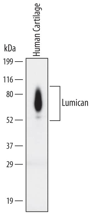
Detection of Human Lumican by Western Blot.
Western blot shows lysates of human cartilage tissue. PVDF Membrane was probed with 2 µg/mL of Human Lumican Monoclonal Antibody (Catalog # MAB28461) followed by HRP-conjugated Anti-Mouse IgG Secondary Antibody (Catalog # HAF007). Differentially glycosylated Lumican protein species were detected between 65 and 85 kDa (as indicated). This experiment was conducted under non-reducing conditions and using Immunoblot Buffer Group 1.Applications for Human/Mouse Lumican Antibody
Western Blot
Sample: Human cartilage tissue
Formulation, Preparation, and Storage
Purification
Reconstitution
Formulation
Shipping
Stability & Storage
- 12 months from date of receipt, -20 to -70 °C as supplied.
- 1 month, 2 to 8 °C under sterile conditions after reconstitution.
- 6 months, -20 to -70 °C under sterile conditions after reconstitution.
Background: Lumican
Lumican is a secreted matrix protein that belongs to the small leucine‑rich proteoglycan (SLRP) class II family. Mature Lumican has a negatively charged N‑terminal domain that contains tyrosine sulfates, followed by twelve leucine‑rich repeats, which are flanked by cysteine loops. Lumican is expressed widely in mammalian connective tissues, while corneal Lumican exists as a keratan sulfate proteoglycan in adult cartilage, Lumican exists primarily as a glycoprotein and lacks keratan sulfate. The amino acid sequence of human Lumican shares 88% amino acid sequence identity with that of mature mouse Lumican.
Alternate Names
Gene Symbol
UniProt
Additional Lumican Products
Product Documents for Human/Mouse Lumican Antibody
Product Specific Notices for Human/Mouse Lumican Antibody
For research use only
