Human/Mouse MAGI2 Antibody
R&D Systems, part of Bio-Techne | Catalog # AF7117

Key Product Details
Species Reactivity
Validated:
Cited:
Applications
Validated:
Cited:
Label
Antibody Source
Product Specifications
Immunogen
Ser2-Arg130
Accession # Q86UL8
Specificity
Clonality
Host
Isotype
Scientific Data Images for Human/Mouse MAGI2 Antibody
Detection of Human MAGI2 by Western Blot.
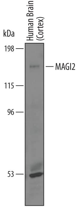
Western blot shows lysates of human brain (cortex) tissue. PVDF membrane was probed with 1 µg/mL of Goat Anti-Human MAGI2 Antigen Affinity-purified Polyclonal Antibody (Catalog # AF7117) followed by HRP-conjugated Anti-Goat IgG Secondary Antibody (Catalog # HAF019). A specific band was detected for MAGI2 at approximately 170 kDa (as indicated). This experiment was conducted under reducing conditions and using Immunoblot Buffer Group 1.MAGI2 in U‑87 MG Human Cell Line.
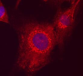
MAGI2 was detected in immersion fixed U-87 MG human glioblastoma/ astrocytoma cell line using Goat Anti-Human MAGI2 Antigen Affinity-purified Polyclonal Antibody (Catalog # AF7117) at 10 µg/mL for 3 hours at room temperature. Cells were stained using the NorthernLights™ 557-conjugated Anti-Goat IgG Secondary Antibody (red; Catalog # NL001) and counterstained with DAPI (blue). Specific staining was localized to cytoplasm. View our protocol for Fluorescent ICC Staining of Cells on Coverslips.MAGI2 in Human Brain.
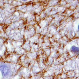
MAGI2 was detected in immersion fixed paraffin-embedded sections of human brain (hippocampus) using Goat Anti-Human MAGI2 Antigen Affinity-purified Polyclonal Antibody (Catalog # AF7117) at 15 µg/mL overnight at 4 °C. Tissue was stained using the Anti-Goat HRP-DAB Cell & Tissue Staining Kit (brown; Catalog # CTS008) and counterstained with hematoxylin (blue). Specific staining was localized to synaptic boutons and neuronal processes. View our protocol for Chromogenic IHC Staining of Paraffin-embedded Tissue Sections.Applications for Human/Mouse MAGI2 Antibody
Immunocytochemistry
Sample: Immersion fixed U‑87 MG human glioblastoma/astrocytoma cell line
Immunohistochemistry
Sample: Immersion fixed paraffin-embedded sections of human brain (hippocampus)
Western Blot
Sample: Human brain (cortex) tissue
Formulation, Preparation, and Storage
Purification
Reconstitution
Formulation
Shipping
Stability & Storage
- 12 months from date of receipt, -20 to -70 °C as supplied.
- 1 month, 2 to 8 °C under sterile conditions after reconstitution.
- 6 months, -20 to -70 °C under sterile conditions after reconstitution.
Background: MAGI2
MAGI2 (AIP-1, ACVRINP1 also known as Activin receptor-interacting protein 1 and S-SCAM in rodents); is a 160‑180 kDa member of the MAGUK family of proteins. It is found in neuronal post-synaptic membrane complexes, and serves as a molecular scaffold for multiple proteins, including alpha-actinin, dendrin, SMAD3 and beta‑catenin. ARIP-1 facilitates the signaling of both growth factor and neurotransmitter receptors such as ActRIIA, NMDA and beta1-adrenergic receptors. Human ARIP-1 is 1455 amino acids (aa) in length. It contains an N-terminal PZD domain (aa 17-101), followed by a guanylate kinase-like domain (aa 109-283), two WW domains (aa 302‑381) and five subsequent PZD domains (aa 426-1229). ARIP-1 is reported to dimerize/oligomerize. There are three potential isoform variants. All utilize an alternative start site at Met164 that may be accompanied by either an Arg substitution for aa 757-771, or a 48 aa substitution for aa 1236-1455. Over aa 2-130, human and mouse ARIP-1 are identical in aa sequence.
Long Name
Alternate Names
Gene Symbol
UniProt
Additional MAGI2 Products
Product Documents for Human/Mouse MAGI2 Antibody
Product Specific Notices for Human/Mouse MAGI2 Antibody
For research use only


