Human/Mouse Phospho-Akt (S473) Pan Specific Antibody
R&D Systems, part of Bio-Techne | Catalog # MAB887


Conjugate
Catalog #
Key Product Details
Validated by
Biological Validation
Species Reactivity
Validated:
Human, Mouse
Cited:
Human, Mouse
Applications
Validated:
Simple Western, Western Blot
Cited:
Flow Cytometry, Infared Microscopy, Western Blot
Label
Unconjugated
Antibody Source
Monoclonal Mouse IgG1 Clone # 545007
Product Specifications
Immunogen
Phosphopeptide containing the human Akt (S473) site
Specificity
Detects human and mouse Akt1, Akt2 and Akt3, when phosphorylated at S473, S474 and S472, respectively.
Clonality
Monoclonal
Host
Mouse
Isotype
IgG1
Scientific Data Images for Human/Mouse Phospho-Akt (S473) Pan Specific Antibody
Detection of Human and Mouse Phospho-Akt (S473) by Western Blot.
Western blot shows lysates of Jurkat human acute T cell leukemia cell line, MCF-7 human breast cancer cell line, and NIH-3T3 mouse embryonic fibroblast cell line untreated (-) or treated (+) with 100 nm Calyculin for 30 minutes, 1 µg/mL insulin for 5 minutes, or 100 ng/mL Recombinant Human PDGF-AB (Catalog # 222-AB) for 20 minutes. PVDF membrane was probed with 0.2 µg/mL of Mouse Anti-Human Phospho-Akt (S473) Pan Specific Monoclonal Antibody (Catalog # MAB887) followed by HRP-conjugated Anti-Mouse IgG Secondary Antibody (Catalog # HAF007). A specific band was detected for Phospho-Akt (S473) at approximately 65 kDa (as indicated). This experiment was conducted under reducing conditions and using Immunoblot Buffer Group 1.Detection of Human Phospho-Akt (S473) by Simple WesternTM.
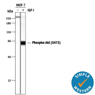
Simple Western lane view shows lysates of MCF-7 human breast cancer cell line untreated (-) or treated (+) with 100 ng/mL Recombinant Human IGF-I (Catalog # 291-G1) for 20 minutes, loaded at 0.2 mg/mL. A specific band was detected for Phospho-Akt (S473) at approximately 65 kDa (as indicated) using 2 µg/mL of Mouse Anti-Human/Mouse Phospho-Akt (S473) Pan Specific Monoclonal Antibody (Catalog # MAB887). This experiment was conducted under reducing conditions and using the 12-230 kDa separation system.Detection of Human AKT by Simple Western
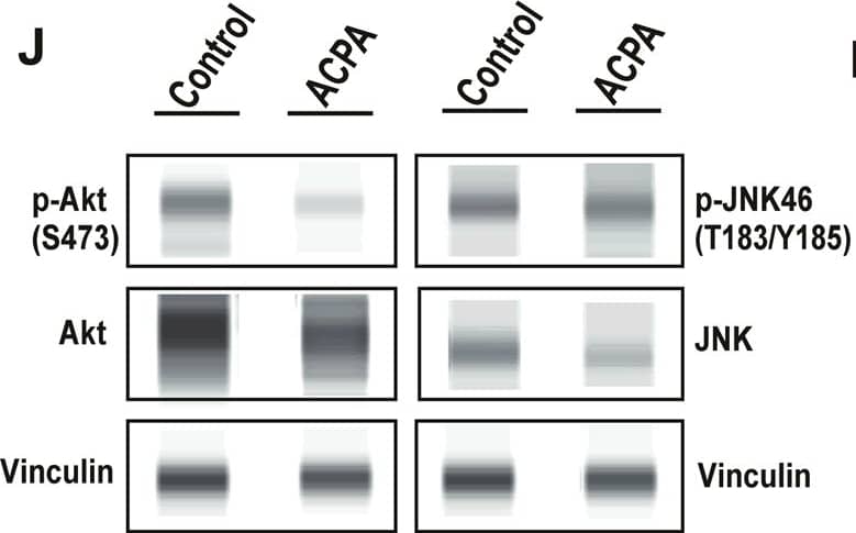
ACPA inhibits p-Akt, induces p-JNK and affects levels of specific metabolites in NSCLC lines.a Principal component analysis (PCA) score plot: Metabolomics profiling of control and ACPA-treated A549, H1299, H358, and H838 cells. b Changes in variable importance in projection (VIP) values for 19 metabolites in A549 cells. c, d, e Changes in VIP values for 20 metabolites in H1299, H358, and H838 cells. Significantly changed metabolites (*p < 0.05, indicated by arrows) were matched to apoptotic pathways. f, g, h, i Increase and decrease in several metabolites of ACPA-treated A549, H1299, H358, and H838 cells (*p < 0.05). j Simple Western showing total Akt, p-Akt (S473), total JNK46 and JNK54 and p-JNK46 and p-JNK54 (T183/Y185) in A549 cells at 24 hours after treatment with IC50 dose of ACPA. k Relative expression levels of Akt and p-Akt for control and ACPA-treated A549 cells after normalization by total vinculin protein. l Relative expression levels of JNK (46 and 54 kDa) and p-JNK for control and ACPA-treated A549 cells after normalization by total vinculin protein. *p < 0.05, Student’s t-test. All tests were done in quadruplicates. Image collected and cropped by CiteAb from the following publication (https://pubmed.ncbi.nlm.nih.gov/33431819), licensed under a CC-BY license. Not internally tested by R&D Systems.Applications for Human/Mouse Phospho-Akt (S473) Pan Specific Antibody
Application
Recommended Usage
Simple Western
2 µg/mL
Sample: MCF‑7 human breast cancer cell line treated with Recombinant Human IGF‑I (Catalog # 291-G1)
Sample: MCF‑7 human breast cancer cell line treated with Recombinant Human IGF‑I (Catalog # 291-G1)
Western Blot
0.2 µg/mL
Sample: Jurkat human acute T cell leukemia cell line, MCF‑7 human breast cancer cell line, and NIH‑3T3 mouse embryonic fibroblast cell line treated with calyculin, insulin, or Recombinant Human PDGF-AB (Catalog # 222-AB)
Sample: Jurkat human acute T cell leukemia cell line, MCF‑7 human breast cancer cell line, and NIH‑3T3 mouse embryonic fibroblast cell line treated with calyculin, insulin, or Recombinant Human PDGF-AB (Catalog # 222-AB)
Reviewed Applications
Read 2 reviews rated 5 using MAB887 in the following applications:
Formulation, Preparation, and Storage
Purification
Protein A or G purified from hybridoma culture supernatant
Reconstitution
Sterile PBS to a final concentration of 0.5 mg/mL. For liquid material, refer to CoA for concentration.
Formulation
Lyophilized from a 0.2 μm filtered solution in PBS with Trehalose. *Small pack size (SP) is supplied either lyophilized or as a 0.2 µm filtered solution in PBS.
Shipping
Lyophilized product is shipped at ambient temperature. Liquid small pack size (-SP) is shipped with polar packs. Upon receipt, store immediately at the temperature recommended below.
Stability & Storage
Use a manual defrost freezer and avoid repeated freeze-thaw cycles.
- 12 months from date of receipt, -20 to -70 °C as supplied.
- 1 month, 2 to 8 °C under sterile conditions after reconstitution.
- 6 months, -20 to -70 °C under sterile conditions after reconstitution.
Background: Akt
Long Name
v-Akt Murine Thymoma Viral Oncogene Homolog
Alternate Names
PKB, RAC
Additional Akt Products
Product Documents for Human/Mouse Phospho-Akt (S473) Pan Specific Antibody
Product Specific Notices for Human/Mouse Phospho-Akt (S473) Pan Specific Antibody
For research use only
Loading...
Loading...
Loading...
Loading...

