Human/Mouse/Rat Cyclophilin B Antibody
R&D Systems, part of Bio-Techne | Catalog # AF5410

Key Product Details
Validated by
Species Reactivity
Applications
Label
Antibody Source
Product Specifications
Immunogen
Asp34-Glu216
Accession # P23284
Specificity
Clonality
Host
Isotype
Scientific Data Images for Human/Mouse/Rat Cyclophilin B Antibody
Detection of Human/Mouse/Rat Cyclophilin B by Western Blot.
Western blot shows lysates of HeLa human cervical epithelial carcinoma cell line, A431 human epithelial carcinoma cell line, BaF3 mouse pro-B cell line, and Y3-Ag rat myeloid cell line. PVDF membrane was probed with 1 µg/mL Goat Anti-Human/Mouse/Rat Cyclophilin B Antigen Affinity-purified Polyclonal Antibody (Catalog # AF5410) followed by HRP-conjugated Anti-Goat IgG Secondary Antibody (Catalog # HAF019). For additional reference, recombinant human Cyclophilin A and Cyclophilin B (5 ng/lane) were included. A specific band for Cyclophilin B was detected at approximately 24 kDa (as indicated). This experiment was conducted under reducing conditions and using Immunoblot Buffer Group 1.Cyclophilin B in HeLa Human Cell Line.
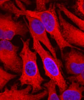
Cyclophilin B was detected in immersion fixed HeLa human cervical epithelial carcinoma cell line using Goat Anti-Human/Mouse/Rat Cyclophilin B Antigen Affinity-purified Polyclonal Antibody (Catalog # AF5410) at 10 µg/mL for 3 hours at room temperature. Cells were stained using the Northern-Lights™ 557-conjugated Anti-Goat IgG Secondary Antibody (red; Catalog # NL001) and counterstained with DAPI (blue). Specific staining was localized to cytoplasm. View our protocol for Fluorescent ICC Staining of Cells on Coverslips.Cyclophilin B in HeLa Human Cell Line.
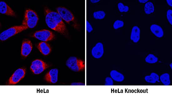
Cyclophilin B was detected in immersion fixed wildtype (left panel) but is not detected in Cyclophilin B knockout (right panel) HeLa human cervical epithelial carcinoma cell line using Goat Anti-Human/Mouse/Rat Cyclophilin B Antigen Affinity-purified Polyclonal Antibody (Catalog # AF5410) at 1 µg/mL for 3 hours at room temperature. Cells were stained using the NorthernLights™ 557-conjugated Anti-Goat IgG Secondary Antibody (red; Catalog # NL001) and counterstained with DAPI (blue). Specific staining was localized to cytoplasm. View our protocol for Fluorescent ICC Staining of Cells on Coverslips.Applications for Human/Mouse/Rat Cyclophilin B Antibody
Immunocytochemistry
Sample: Immersion fixed HeLa human cervical epithelial carcinoma cell line
Knockout Validated
Western Blot
Sample: HeLa human cervical epithelial carcinoma cell line, A431 human epithelial carcinoma cell line, BaF3 mouse pro-B cell line, and Y3-Ag rat myeloid cell line
Formulation, Preparation, and Storage
Purification
Reconstitution
Formulation
Shipping
Stability & Storage
- 12 months from date of receipt, -20 to -70 °C as supplied.
- 1 month, 2 to 8 °C under sterile conditions after reconstitution.
- 6 months, -20 to -70 °C under sterile conditions after reconstitution.
Background: Cyclophilin B
Cyclophilin B (SCYLP, CyPB and peptidyl-prolyl cis-trans isomerase B) is a 24 kDa glycoprotein member of the B subfamily of the cyclophilin-type PPIase family of molecules. It is both secreted and retained in the ER. When secreted, it mediates chemotaxis and T cell adhesion to fibronectin. This is likely due to its prolyl cis/trans isomerase activity. Intracellularly, Cyclophilin B appears to serve as a molecular chaperone for molecules destined for secretion. It does so via stabilization, and facilitating the activity of additional chaperones. The human CyPB precursor is 216 amino acids (aa) in length. It contains a 25 aa signal sequence plus a 191 aa mature region. There is a partial heparin-binding sequence (aa 27‑34), a PPIase domain (aa 47‑204) and a C-terminal ER retention motif (aa 213‑216). Over aa 34‑216, the human and mouse sequences are 95% aa identical.
Alternate Names
Gene Symbol
UniProt
Additional Cyclophilin B Products
Product Documents for Human/Mouse/Rat Cyclophilin B Antibody
Product Specific Notices for Human/Mouse/Rat Cyclophilin B Antibody
For research use only


