Human/Mouse/Rat Jak1 Antibody
R&D Systems, part of Bio-Techne | Catalog # MAB4260


Key Product Details
Species Reactivity
Validated:
Cited:
Applications
Validated:
Cited:
Label
Antibody Source
Product Specifications
Immunogen
Pro32-Phe286
Accession # P23458
Specificity
Clonality
Host
Isotype
Scientific Data Images for Human/Mouse/Rat Jak1 Antibody
Detection of Jak1 in Jurkat Human Cell Line by Flow Cytometry.
Jurkat human acute T cell leukemia cell line was stained with Rat Anti-Human/Mouse/Rat Jak1 Monoclonal Antibody (Catalog # MAB4260, filled histogram) or isotype control antibody (MAB0061, open histogram), followed by Phycoerythrin-conjugated Anti-Rat IgG F(ab')2Secondary Antibody (F0105B). To facilitate intracellular staining, cells were fixed with paraformaldehyde and permeabilized with saponin.Detection of Human and Mouse Jak1 by Western Blot.
Western blot shows lysates of Jurkat human acute T cell leukemia cell line, K562 human chronic myelogenous leukemia cell line, A20 mouse B cell lymphoma cell line, and L1.2 mouse pro-B cell line. PVDF membrane was probed with 1 µg/mL of Rat Anti-Human/Mouse/Rat Jak1 Monoclonal Antibody (Catalog # MAB4260) followed by HRP-conjugated Anti-Rat IgG Secondary Antibody (Catalog # HAF005). A specific band was detected for Jak1 at approximately 130 kDa (as indicated). This experiment was conducted under reducing conditions and using Immunoblot Buffer Group 3.Jak1 in Human Epidermis.
Jak1 was detected in immersion fixed paraffin-embedded sections of human epidermis using 25 µg/mL Rat Anti-Human/Mouse/Rat Jak1 Monoclonal Antibody (Catalog # MAB4260) overnight at 4 °C. Tissue was stained with the Anti-Rat HRP-DAB Cell & Tissue Staining Kit (brown; CTS017) and counterstained with hematoxylin (blue). View our protocol for Chromogenic IHC Staining of Paraffin-embedded Tissue Sections.Applications for Human/Mouse/Rat Jak1 Antibody
CyTOF-ready
Immunocytochemistry
Sample: Immersion fixed HeLa human cervical epithelial carcinoma cell line
Immunohistochemistry
Sample: Immersion fixed paraffin-embedded sections of human epidermis
Intracellular Staining by Flow Cytometry
Sample: Jurkat human acute T cell leukemia cell line fixed with paraformaldehyde and permeabilized with saponin
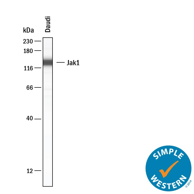
Simple Western
Sample: Lysates of Daudi human Burkitt's lymphoma cell line.
Western Blot
Sample: Jurkat human acute T cell leukemia cell line, K562 human chronic myelogenous leukemia cell line, A20 mouse B cell lymphoma cell line, and L1.2 mouse pro-B cell line
Reviewed Applications
Read 4 reviews rated 5 using MAB4260 in the following applications:
Formulation, Preparation, and Storage
Purification
Reconstitution
Formulation
Shipping
Stability & Storage
- 12 months from date of receipt, -20 to -70 °C as supplied.
- 1 month, 2 to 8 °C under sterile conditions after reconstitution.
- 6 months, -20 to -70 °C under sterile conditions after reconstitution.
Background: Jak1
Janus Kinase 1 (Jak1) belongs to a family of protein tyrosine kinases that couple to cytokine receptors and are activated by ligand binding to these receptors. Activation of Jak1 occurs via phosphorylation at two adjacent tyrosine residues, Y1022 and Y1023, within the kinase domain. Jaks activate members of the STAT family of transcription factors by phosphorylating critical tyrosine regulatory sites. Jak1 is required for the activation of STAT1 and STAT2 in response to interferon alpha.
Long Name
Alternate Names
Entrez Gene IDs
Gene Symbol
UniProt
Additional Jak1 Products
Product Documents for Human/Mouse/Rat Jak1 Antibody
Product Specific Notices for Human/Mouse/Rat Jak1 Antibody
This product is sold under license from Millipore Corporation under the following US or foreign patents: 5,821,069; 5,658,791; EP0560890. This product shall not be used to commercially screen drug molecules being developed as JAK1 or JAK2 inhibitors. Any such activity will be outside the scope of the research use only label license.
For research use only


