Human/Mouse/Rat Phospho-CDC2/CDK1 (Y15) Antibody
R&D Systems, part of Bio-Techne | Catalog # AF888


Discontinued Product
AF888 has been discontinued.
View all CDC2/CDK1 products.
Key Product Details
Validated by
Biological Validation
Species Reactivity
Validated:
Human, Mouse, Rat
Cited:
Mouse, Rat
Applications
Validated:
Immunocytochemistry, Western Blot
Cited:
Neutralization, Western Blot
Label
Unconjugated
Antibody Source
Polyclonal Rabbit IgG
Product Specifications
Immunogen
Phosphopeptide containing CDC2 Y15 site
Specificity
Detects human, mouse, and rat CDC2 when phosphorylated at Y15.
Clonality
Polyclonal
Host
Rabbit
Isotype
IgG
Scientific Data Images for Human/Mouse/Rat Phospho-CDC2/CDK1 (Y15) Antibody
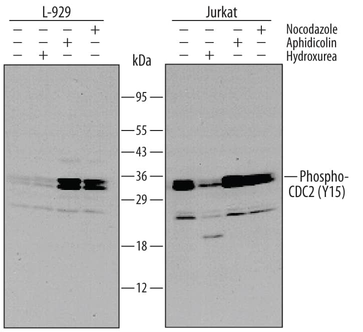
Detection of Mouse and Human Phospho-CDC2 (Y15) by Western Blot.
Western blot shows lysates of L-929 mouse fibroblast cell line and Jurkat human acute T cell leukemia cell line untreated (-) or treated (+) with 0.2 µg/mL nocodazole or 12 µM aphidicolin or 1 mM hydroxurea for 18 hours. PVDF membrane was probed with 0.2 µg/mL of Rabbit Anti-Human/Mouse/Rat Phospho-CDC2 (Y15) Antigen Affinity-purified Polyclonal Antibody (Catalog # AF888), followed by HRP-conjugated Anti-Rabbit IgG Secondary Antibody (Catalog # HAF008). A specific band was detected for Phospho-CDC2 (Y15) at approximately 34 kDa (as indicated). This experiment was conducted under reducing conditions and using Immunoblot Buffer Group 1.Detection of Human CDC2/CDK1 by Western Blot
Cooperative induction of DNA damage in vivo by WEE1 and CHK1 inhibitors. LoVo xenograft tumor-bearing mice were treated with 60 mpk MK-1775 BID for 2 days, 60 mpk MK-8776 BID for 2 days, or the combination of MK-1775 and MK-8776 each at 60 mpk BID for 2 days. Tumors were collected at 2, 24, and 48 hours following the final dose. A, LoVo tumor lysates were analyzed by Western blot for pCHK1S345. B, Tumor sections were fixed and analyzed by immunohistochemistry (IHC). Representative images for gammaH2AX at 2 hours and 48 hours post final dose are shown. C, Quantitative analysis of IHC for both phospho-CHK1S345 and gammaH2AX (n=3); one-way ANOVA analyses *P<0.05. **P<0.01, ***P<0.001. Image collected and cropped by CiteAb from the following open publication (https://pubmed.ncbi.nlm.nih.gov/23148684), licensed under a CC-BY license. Not internally tested by R&D Systems.Detection of Human CDC2/CDK1 by Western Blot
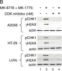
DNA damage response incurred by MK-1775 and MK-8776 is dependent on CDK activity.A, Resistant (H460) or sensitive (LoVo) cells were treated with concentrations of MK-1775 and MK-8776 described for Figure \n4, or 1 uM nocodazole for control. After 24 hours, cells were harvested and lysates analyzed by Western blot for caspase-dependent cleaved PARP (PARP*). B, A2058, HT-29, and LoVo cells were treated for 30 minutes with either DMSO or the indicated concentration of CDK inhibitor (SCH-727965). Following this pretreatment, further DMSO or concentrations of MK-1775 and MK-8776 used in Figures \n3 and\n4 (125 nM MK-1775 plus 150 nM MK-8776 in A2058; 125 nM MK-1775 plus 300 nM MK-8776 in HT-29, and 40 nM MK-1775 plus 75 nM MK-8776 in LoVo) were added to the cells for an additional 2 hours before cells were harvested and lysates analyzed by Western blot for phosphorylated CHK1S345, indicative of activated DNA damage response. C, LoVo cells were treated for 2 hours with 75 nM MK-1775 alone or in combination with 150 nM MK-8776, as indicated. Cells were harvested and lysates analyzed by Western blot for the proteins and phosphoproteins indicated. Image collected and cropped by CiteAb from the following open publication (https://pubmed.ncbi.nlm.nih.gov/23148684), licensed under a CC-BY license. Not internally tested by R&D Systems.Applications for Human/Mouse/Rat Phospho-CDC2/CDK1 (Y15) Antibody
Application
Recommended Usage
Immunocytochemistry
5-15 µg/mL
Sample: Immersion fixed MCF-7 human breast cancer cell line
Sample: Immersion fixed MCF-7 human breast cancer cell line
Western Blot
0.2 µg/mL
Sample: Jurkat human acute T cell leukemia cell line
Sample: Jurkat human acute T cell leukemia cell line
Reviewed Applications
Read 4 reviews rated 3.8 using AF888 in the following applications:
Formulation, Preparation, and Storage
Purification
Antigen Affinity-purified
Reconstitution
Reconstitute at 0.2 mg/mL in sterile PBS. For liquid material, refer to CoA for concentration.
Formulation
Lyophilized from a 0.2 μm filtered solution in PBS with Trehalose. *Small pack size (SP) is supplied either lyophilized or as a 0.2 µm filtered solution in PBS.
Shipping
Lyophilized product is shipped at ambient temperature. Liquid small pack size (-SP) is shipped with polar packs. Upon receipt, store immediately at the temperature recommended below.
Stability & Storage
Use a manual defrost freezer and avoid repeated freeze-thaw cycles.
- 12 months from date of receipt, -20 to -70 °C as supplied.
- 1 month, 2 to 8 °C under sterile conditions after reconstitution.
- 6 months, -20 to -70 °C under sterile conditions after reconstitution.
Background: CDC2/CDK1
Long Name
Cell Division Cycle 2/Cyclin-dependent Kinase 1
Alternate Names
CDK1
Gene Symbol
CDK1
Additional CDC2/CDK1 Products
Product Documents for Human/Mouse/Rat Phospho-CDC2/CDK1 (Y15) Antibody
Product Specific Notices for Human/Mouse/Rat Phospho-CDC2/CDK1 (Y15) Antibody
For research use only
Loading...
Loading...
Loading...
Loading...


