Human/Mouse/Rat Relaxin R1 Antibody
R&D Systems, part of Bio-Techne | Catalog # MAB8898


Key Product Details
Species Reactivity
Validated:
Cited:
Applications
Validated:
Cited:
Label
Antibody Source
Product Specifications
Immunogen
Met1-Ser398
Accession # Q9HBX9
Specificity
Clonality
Host
Isotype
Scientific Data Images for Human/Mouse/Rat Relaxin R1 Antibody
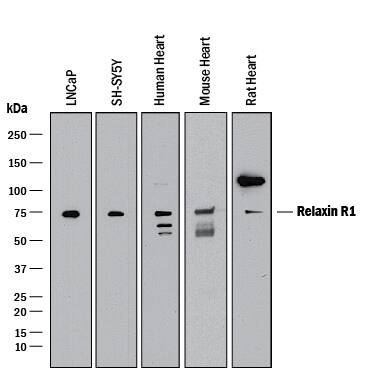
Detection of Human, Mouse, and Rat Relaxin R1 by Western Blot.
Western blot shows lysates of LNCaP human prostate cancer cell line, SH-SY5Y human neuroblastoma cell line, human heart tissue, mouse heart tissue, and rat heart tissue. PVDF membrane was probed with 2 µg/mL of Mouse Anti-Human Relaxin R1 Monoclonal Antibody (Catalog # MAB8898) followed by HRP-conjugated Anti-Mouse IgG Secondary Antibody (Catalog # HAF018). A specific band was detected for Relaxin R1 at approximately 75 kDa (as indicated). This experiment was conducted under reducing conditions and using Immunoblot Buffer Group 1.Relaxin R1 in Mouse Brain.
Relaxin R1 was detected in immersion fixed frozen sections of mouse brain (medulla) using Mouse Anti-Human Relaxin R1 Monoclonal Antibody (Catalog # MAB8898) at 2 µg/mL overnight at 4 °C. Tissue was stained using the NorthernLights™ 557-conjugated Anti-Mouse IgG Secondary Antibody (red; Catalog # NL007) and counterstained with DAPI (blue). Specific staining was localized to neurons. View our protocol for Fluorescent IHC Staining of Frozen Tissue Sections.Relaxin R1 in Human Prostate.
Relaxin R1 was detected in immersion fixed paraffin-embedded sections of human prostate using Mouse Anti-Human/Mouse/Rat Relaxin R1 Monoclonal Antibody (Catalog # MAB8898) at 1 µg/mL for 1 hour at room temperature followed by incubation with the Anti-Mouse IgG VisUCyte™ HRP Polymer Antibody (VC001). Before incubation with the primary antibody, tissue was subjected to heat-induced epitope retrieval using Antigen Retrieval Reagent-Basic (CTS013). Tissue was stained using DAB (brown) and counterstained with hematoxylin (blue). Specific staining was localized to epithelial cells in prostate glands. Staining was performed using our protocol for IHC Staining with VisUCyte HRP Polymer Detection Reagents.Applications for Human/Mouse/Rat Relaxin R1 Antibody
CyTOF-ready
Flow Cytometry
Sample: SH-SY5Y human neuroblastoma cell line
Immunohistochemistry
Sample: Immersion fixed frozen sections of mouse brain (medulla) and immersion fixed paraffin-embedded sections of human prostate
Western Blot
Sample: LNCaP human prostate cancer cell line, SH‑SY5Y human neuroblastoma cell line, human heart tissue, mouse heart tissue, and rat heart tissue
Formulation, Preparation, and Storage
Purification
Reconstitution
Formulation
Shipping
Stability & Storage
- 12 months from date of receipt, -20 to -70 °C as supplied.
- 1 month, 2 to 8 °C under sterile conditions after reconstitution.
- 6 months, -20 to -70 °C under sterile conditions after reconstitution.
Background: Relaxin R1
Relaxin R1 (Relaxin Receptor 1), also known as RXFP1 (Relaxin Family Peptide Receptor 1) or LGR7 (Leucine‑rich G‑protein‑coupled Receptor 7) is a member of family C of the LGRs, and is one of four receptors for Relaxin family proteins. Relaxin R1 shows highest affinity for human Relaxins 1, 2 and 3, while RXFP2 binds Relaxin 2 and the related INSL3, and RXFP3 primarily binds Relaxin 3 (1, 2). The 757 amino acid (aa) human Relaxin R1 contains an N‑terminal 409 aa extracellular domain (ECD) with a calcium‑binding LDL R class A (LDLa) domain and 10 leucine‑rich repeats (LRR) with several N‑glycosylation sites. The C‑terminus contains 12 transmembrane domains within aa 410‑672. Human Relaxin R1 (aa 1‑398) shares 84, 86, 85, 85 and 91% aa sequence identity with mouse, rat, equine, bovine and porcine Relaxin R1, respectively. Isoforms of 724 and 709 aa lack aa 63‑96 and 300‑348, respectively, while isoforms of 176, 189, 191 and 337 aa diverge after aa 154, 179, 181 and 324, respectively (3, 4). These forms may dimerize with full‑length Relaxin R1 and reduce its expression on the cell surface (3, 4). Receptor activation and cAMP signaling depend on the LDLa domain, and Relaxin binding requires the LRR repeats, with a secondary binding site within transmembrane region exoloops (1, 2, 5). Of LGR family members, RXFP1 and RXFP2 are unique in that they are not internalized to down‑regulate signaling, and their LDLa domains allow transmission of both G‑protein‑dependent and ‑independent signals (1, 2, 6, 7). Engagement of Relaxin R1 by Relaxin (mainly Relaxin 2 in humans) supports female reproduction by promoting uterine angiogenesis, ovarian follicle ripening, and endometrial, cervical and nipple development (8‑10). In male reproduction, Relaxin R1 acts in the prostate to enhance sperm motility (11). It reduces fibrosis in the heart, skin, lungs, liver, kidney, and reproductive tissues by combating aberrant collagen buildup (12). In the vasculature, it mediates vasodilation and decreases blood pressure. Relaxin R1 is expressed on human leukocytes and promotes adhesion, migration, and osteoclast differentiation (13, 14). Additional effects on heart, lungs, kidney and brain are reported, some of which may be species‑specific (1).
References
- van der Westhuizen, E.T. et al. (2008) Drug Discov. Today 13:640.
- Kong, R.C.K. et al. (2010) Mol. Cell. Endocrinol. 320:1.
- Muda, M. et al. (2005) Mol. Hum. Reprod. 11:591.
- Kern, A. et al. (2008) Endocrinology 149:1227.
- Hopkins, E.J. et al. (2007) J. Biol. Chem. 282:4172.
- Kern, A. and G.D. Bryant-Greenwood (2009) Endocrinology 150:2419.
- Halls, M.L. (2012) Br. J. Pharmacol. 165:1644.
- Kamat, A.A. et al. (2004) Endocrinology 145:4712.
- Krajnc-Franken, M.A. et al. (2004) Mol. Cell. Biol. 24:687.
- Yao, L. et al. (2008) Endocrinology 149:2072.
- Ferlin, A. et al. (2012) J. Androl. 33:474.
- Hossain, M.A. (2011) Biochemistry 50:1368.
- Ferlin, A. et al. (2010) Bone 46:504.
- Figueiredo, K.A. et al. (2006) J. Biol. Chem. 281:3030.
Long Name
Alternate Names
Gene Symbol
UniProt
Additional Relaxin R1 Products
Product Documents for Human/Mouse/Rat Relaxin R1 Antibody
Product Specific Notices for Human/Mouse/Rat Relaxin R1 Antibody
For research use only


