Human Myeloperoxidase/MPO Antibody
R&D Systems, part of Bio-Techne | Catalog # AF3174


Key Product Details
Validated by
Species Reactivity
Validated:
Cited:
Applications
Validated:
Cited:
Label
Antibody Source
Product Specifications
Immunogen
Ala49-Ser745
Accession # P05164
Specificity
Clonality
Host
Isotype
Scientific Data Images for Human Myeloperoxidase/MPO Antibody
Detection of Human Myeloperoxidase/MPO by Western Blot.
Western blot shows lysates of HL-60 human acute promyelocytic leukemia cell line and human neutrophil. PVDF membrane was probed with 0.5 µg/mL of Goat Anti-Human Myeloperoxidase/MPO Antigen Affinity-purified Polyclonal Antibody (Catalog # AF3174) followed by HRP-conjugated Anti-Goat IgG Secondary Antibody (Catalog # HAF019). A specific band was detected for Myeloperoxidase/MPO at approximately 60-65 kDa (as indicated). This experiment was conducted under reducing conditions and using Immunoblot Buffer Group 2.Myeloperoxidase/MPO in HL-60 Human Cell Line.
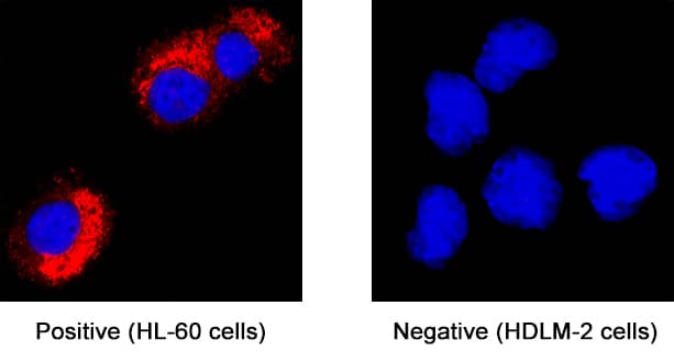
Myeloperoxidase/MPO was detected in immersion fixed HL-60 human acute promyelocytic leukemia cell line (positive staining) and HDLM-2 human Hodgkin’s lymphoma cell line (negative staining) using Goat Anti-Human Myeloperoxidase/MPO Antigen Affinity-purified Polyclonal Antibody (Catalog # AF3174) at 1.7 µg/mL for 3 hours at room temperature. Cells were stained using the NorthernLights™ 557-conjugated Anti-Goat IgG Secondary Antibody (red; NL001) and counterstained with DAPI (blue). Specific staining was localized to cytoplasm. Staining was performed using our protocol for Fluorescent ICC Staining of Non-adherent Cells.Detection of Human Myeloperoxidase/MPO by Simple WesternTM.
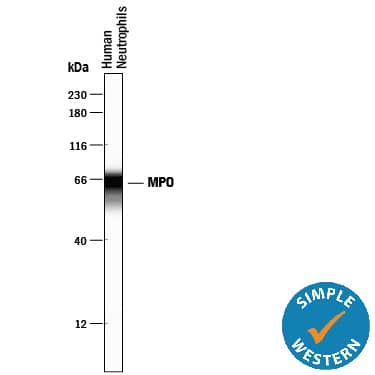
Simple Western lane view shows lysates of human neutrophils, loaded at 0.2 mg/mL. A specific band was detected for Myeloperoxidase/MPO at approximately 65 kDa (as indicated) using 5 µg/mL of Goat Anti-Human Myeloperoxidase/MPO Antigen Affinity-purified Polyclonal Antibody (Catalog # AF3174) followed by 1:50 dilution of HRP-conjugated Anti-Goat IgG Secondary Antibody (Catalog # HAF109). This experiment was conducted under reducing conditions and using the 12-230 kDa separation system.Applications for Human Myeloperoxidase/MPO Antibody
Immunocytochemistry
Sample: Immersion fixed HL‑60 human acute promyelocytic leukemia cell line
Simple Western
Sample: Human neutrophils
Western Blot
Sample: HL‑60 human acute promyelocytic leukemia cell line and human neutrophils
Reviewed Applications
Read 1 review rated 3 using AF3174 in the following applications:
Formulation, Preparation, and Storage
Purification
Reconstitution
Formulation
Shipping
Stability & Storage
- 12 months from date of receipt, -20 to -70 °C as supplied.
- 1 month, 2 to 8 °C under sterile conditions after reconstitution.
- 6 months, -20 to -70 °C under sterile conditions after reconstitution.
Background: Myeloperoxidase/MPO
Myeloperoxidase (MPO) is a heme-containing enzyme belonging to the XPO subfamily of peroxidases. It is an abundant neutrophil and monocyte glycoprotein that catalyzes the hydrogen peroxide-dependent conversion of chloride, bromide, and iodide to multiple reactive species (1). Post-translational processing of MPO involves the insertion of a heme moiety and the proteolytic removal of both a propeptide and a 6 aa internal peptide (2). This results in a disulfide-linked dimer composed of a 60 kDa heavy and 12 kDa light chain that associate into a 150 kDa enzymatically active tetramer. The tetramer contains two heme groups and one disulfide bond between the heavy chains (2). Alternate splicing generates two additional isoforms of MPO, one with a 32 aa insertion in the light chain, and another with a deletion of the signal sequence and part of the propeptide (3). Human and mouse MPO share 87% aa sequence identity. MPO activity results in protein nitrosylation and the formation of 3-chlorotyrosine and dityrosine crosslinks (4‑6). Modification of ApoB100, as well as the lipid and cholesterol components of LDL and HDL, promotes the development of atherosclerosis (5, 7‑9). MPO is also associated with a variety of other diseases (1), and inhibits vasodilation in inflammation by depleting the levels of NO (10). Serum albumin functions as a carrier protein during MPO movement to the basolateral side of epithelial cells (11). MPO is stored in neutrophil azurophilic granules. Upon cellular activation, it is deposited into pathogen-containing phagosomes (2). While mice lacking MPO are impaired in clearing select microbial infections, MPO deficiency in humans does not necessarily result in heightened susceptibility to infections (12, 13).
References
- Klebanoff, S.J. (2005) J. Leukoc. Biol. 77:598.
- Hansson, M. et al. (2006) Arch. Biochem. Biophys. 445:214.
- Hashinaka, K. et al. (1988) Biochemistry 27:5906.
- van Dalen, C.J. et al. (2000) J. Biol. Chem. 275:11638.
- Hazen, S.L. and J.W. Heinecke (1997) J. Clin. Invest. 99:2075.
- Heinecke, J.W. et al. (1993) J. Clin. Invest. 91:2866.
- Podrez, E.A. et al. (1999) J. Clin. Invest. 103:1547.
- Bergt, C. et al. (2004) Proc. Natl. Acad. Sci. 101:13032.
- Hazen, S.L. et al. (1996) J. Biol. Chem. 271:23080.
- Eiserich, J.P. et al. (2002) Science 296:2391.
- Tiruppathi, C. et al. (2004) Proc. Natl. Acad. Sci. 101:7699.
- Aratani Y. et al. (2000) J. Infect. Dis. 182:1276.
- Kutter, D. (1998) J. Mol. Med. 76:669.
Alternate Names
Gene Symbol
UniProt
Additional Myeloperoxidase/MPO Products
Product Documents for Human Myeloperoxidase/MPO Antibody
Product Specific Notices for Human Myeloperoxidase/MPO Antibody
For research use only
