Human p38 delta Antibody
R&D Systems, part of Bio-Techne | Catalog # AF1519

Key Product Details
Species Reactivity
Validated:
Cited:
Applications
Validated:
Cited:
Label
Antibody Source
Product Specifications
Immunogen
Accession # O15264
Specificity
Clonality
Host
Isotype
Scientific Data Images for Human p38 delta Antibody
Detection of Human p38 delta by Western Blot.
Western blot shows lysates of HEK293 human embryonic kidney cell line and HepG2 human hepatocellular carcinoma cell line. PVDF membrane was probed with 0.1 µg/mL Rabbit Anti-Human p38 delta Antigen Affinity-purified Polyclonal Antibody (Catalog # AF1519) followed by HRP-conjugated Anti-Rabbit IgG Secondary Antibody (Catalog # HAF008). For additional reference, recombinant p38 beta, p38 gamma, p38 delta, and p38 alpha at 2 ng/lane) were included. A specific band for p38 delta was detected at approximately 42 kDa (as indicated). This experiment was conducted under reducing conditions and using Immunoblot Buffer Group 1.p38 delta in Human Skin.
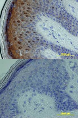
p38d was detected in immersion fixed paraffin-embedded sections of human skin using 15 µg/mL Rabbit Anti-Human p38d Antigen Affinity-purified Polyclonal Antibody (Catalog # AF1519) overnight at 4 °C. Tissue was stained with the Anti-Rabbit HRP-DAB Cell & Tissue Staining Kit (brown; Catalog # CTS005) and counterstained with hematoxylin (blue). View our protocol for Chromogenic IHC Staining of Paraffin-embedded Tissue Sections.p38 delta in Human Skin.
p38d was detected in immersion fixed paraffin-embedded sections of human skin using Rabbit Anti-Human p38d Antigen Affinity-purified Polyclonal Antibody (Catalog # AF1519) at 15 µg/mL overnight at 4 °C. Tissue was stained using the Anti-Rabbit HRP-DAB Cell & Tissue Staining Kit (brown; Catalog # CTS005) and counterstained with hematoxylin (blue). Lower panel shows a lack of labeling if primary antibodies are omitted and tissue is stained only with secondary antibody followed by incubation with detection reagents. View our protocol for Chromogenic IHC Staining of Paraffin-embedded Tissue Sections.Applications for Human p38 delta Antibody
Immunohistochemistry
Sample: Immersion fixed paraffin-embedded sections of human skin
Western Blot
Sample: HEK293 human embryonic kidney cell line and HepG2 human hepatocellular carcinoma cell line
Reviewed Applications
Read 1 review rated 5 using AF1519 in the following applications:
Formulation, Preparation, and Storage
Purification
Reconstitution
Formulation
Shipping
Stability & Storage
- 12 months from date of receipt, -20 to -70 °C as supplied.
- 1 month, 2 to 8 °C under sterile conditions after reconstitution.
- 6 months, -20 to -70 °C under sterile conditions after reconstitution.
Background: p38 delta
The p38 Mitogen-activated Protein Kinases (MAPKs) are a family of four related Ser/Thr kinases activated by proinflammatory cytokines and environmental stresses. All four p38 family members, alpha, beta, gamma, and delta, are phosphorylated by MKK3 and/or MKK6 at dual Thr and Tyr positions within the phosphoacceptor sequence Thr-Gly-Tyr. Once activated, p38 phosphorylates a number of targets, including the nuclear transcription factors ATF2 and Max.
The most frequently analyzed family member, p38 alpha, also known as SAPK2a and MAPK14, was initially purified as a kinase critical to the signaling cascade linking IL-1 to MAPKAPK-2 and the small heat shock protein HSP27. Ubiquitously expressed, p38 alpha is dually phosphorylated by MKK3 and MKK6 at Thr180 and Tyr182. Once activated, p38 alpha phosphorylates a number of targets, including the cytoplasmic kinases MNK 4 and PRAK5 and the nuclear transcription factors ATF2 1 and STAT1. Several promising compounds that inhibit p38 alpha are being investigated as potential therapies for arthritic and inflammatory diseases.
Additional p38 delta Products
Product Documents for Human p38 delta Antibody
Product Specific Notices for Human p38 delta Antibody
For research use only


