Human PD-1 Antibody
R&D Systems, part of Bio-Techne | Catalog # MAB10864

Key Product Details
Species Reactivity
Human
Applications
Blockade of Receptor-ligand Interaction, ELISA, Immunohistochemistry, Western Blot
Label
Unconjugated
Antibody Source
Monoclonal Mouse IgG1 Clone # 1015846
Product Specifications
Immunogen
Human embryonic kidney cell HEK293-derived human PD-1
Leu25-Thr168
Accession # Q15116
Leu25-Thr168
Accession # Q15116
Specificity
Detects human PD-1 in direct ELISAs.
Clonality
Monoclonal
Host
Mouse
Isotype
IgG1
Scientific Data Images for Human PD-1 Antibody
Detection of Human PD‑1 by Western Blot.
Western blot shows lysates of human tonsil tissue. PVDF membrane was probed with 2 µg/mL of Mouse Anti-Human PD-1 Monoclonal Antibody (Catalog # MAB10864) followed by HRP-conjugated Anti-Mouse IgG Secondary Antibody (Catalog # HAF018). A specific band was detected for PD-1 at approximately 45 kDa (as indicated). This experiment was conducted under reducing conditions and using Immunoblot Buffer Group 1.PD‑1 in Human Tonsil.
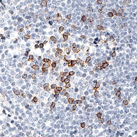
PD-1 was detected in immersion fixed paraffin-embedded sections of human tonsil using Mouse Anti-Human PD-1 Monoclonal Antibody (Catalog # MAB10864) at 5 µg/mL for 1 hour at room temperature followed by incubation with the Anti-Mouse IgG VisUCyte™ HRP Polymer Antibody (Catalog # VC001). Before incubation with the primary antibody, tissue was subjected to heat-induced epitope retrieval using Antigen Retrieval Reagent-Basic (Catalog # CTS013). Tissue was stained using DAB (brown) and counterstained with hematoxylin (blue). Specific staining was localized to lymphocytes. View our protocol for IHC Staining with VisUCyte HRP Polymer Detection Reagents.PD-L1/B7-H1 Binding to PD-1 Blocked by Human PD-1 Antibody.
In a functional ELISA, 0.09-0.72 µg/mL of this antibody (green line) will block 50% of the binding of 5 µg/mL of Recombinant Human PD-L1/B7-H1 Fc Chimera (orange line, Catalog # 156-B7) to immobilized Recombinant Human PD-1 His-tagged Protein (Catalog # 8986-PD) coated at 1 µg/mL (100 µL/well). At 5 µg/mL, this antibody will block >90% of the binding.Applications for Human PD-1 Antibody
Application
Recommended Usage
Blockade of Receptor-ligand Interaction
In a functional ELISA, 0.09-0.72 µg/mL of this antibody will block 50% of the binding of 5 µg/mL of Recombinant Human PD-L1/B7-H1 Fc Chimera (Catalog # 156-B7) to immobilized Recombinant Human PD‑1 His-tagged Protein (Catalog # 8986-PD) coated at 1 µg/mL (100 µL/well). At 5 µg/mL, this antibody will block >90% of the binding.
ELISA
This antibody functions as an ELISA detection antibody when paired with Rabbit Anti-Human PD‑1 Monoclonal Antibody (Catalog # MAB10863).
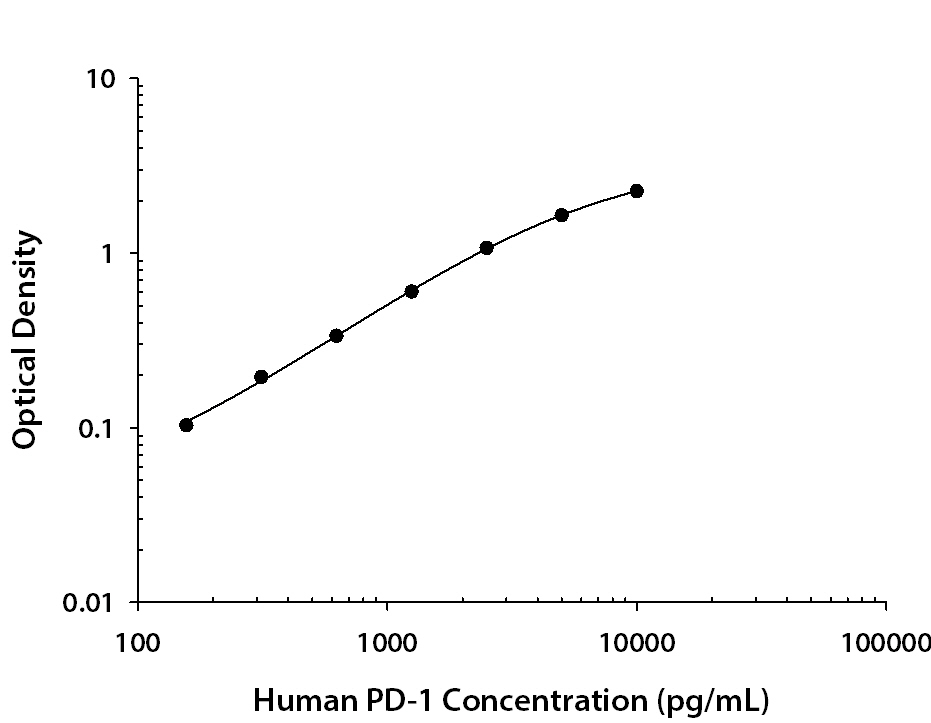
This product is intended for assay development on various assay platforms requiring antibody pairs. We recommend the Human PD-1 DuoSet ELISA Kit (Catalog # DY1086) for convenient development of a sandwich ELISA.
Immunohistochemistry
5-25 µg/mL
Sample: Immersion fixed paraffin-embedded sections of human tonsil
Sample: Immersion fixed paraffin-embedded sections of human tonsil
Western Blot
2 µg/mL
Sample: Human Tonsil Tissue
Sample: Human Tonsil Tissue
Formulation, Preparation, and Storage
Purification
Protein A or G purified from cell culture supernatant
Reconstitution
Reconstitute at 0.5 mg/mL in sterile PBS. For liquid material, refer to CoA for concentration.
Formulation
Lyophilized from a 0.2 μm filtered solution in PBS with Trehalose. *Small pack size (SP) is supplied either lyophilized or as a 0.2 µm filtered solution in PBS.
Shipping
Lyophilized product is shipped at ambient temperature. Liquid small pack size (-SP) is shipped with polar packs. Upon receipt, store immediately at the temperature recommended below.
Stability & Storage
Use a manual defrost freezer and avoid repeated freeze-thaw cycles.
- 12 months from date of receipt, -20 to -70 °C as supplied.
- 1 month, 2 to 8 °C under sterile conditions after reconstitution.
- 6 months, -20 to -70 °C under sterile conditions after reconstitution.
Background: PD-1
References
- Ishida, Y. et al. (1992) EMBO J. 11:3887.
- Sharpe, A.H. and G. J. Freeman (2002) Nat. Rev. Immunol. 2:116.
- Coyle, A. and J. Gutierrez-Ramos (2001) Nat. Immunol. 2:203.
- Nishimura, H. and T. Honjo (2001) Trends Immunol. 22:265.
- Watanabe, N et al. (2003) Nat. Immunol. 4:670.
- Zhang, X. et al. (2004) Immunity 20:337.
- Lázár-Molnár, E. et al. (2008) Proc. Natl. Acad. Sci. USA 105:10483.
- Nishimura, H et al. (1996) Int. Immunol. 8:773.
- Keir, M.E. et al. (2008) Annu. Rev. Immunol. 26:677.
- Butte, M.J. et al. (2007) Immunity 27:111.
- Okazaki, T. et al. (2013) Nat. Immunol. 14:1212.
- Iwai, Y. et al. (2002) Proc. Natl. Acad. Sci. USA 99: 12293.
- Nogrady, B. (2014) Nature 513:S10.
- Swaika, A. et al. (2015) Mol. Immunol. 67: 4
Long Name
Programmed Death-1
Alternate Names
CD279, PD1, PDCD1, SLEB2
Entrez Gene IDs
Gene Symbol
PDCD1
UniProt
Additional PD-1 Products
Product Documents for Human PD-1 Antibody
Product Specific Notices for Human PD-1 Antibody
For research use only
Loading...
Loading...
Loading...
Loading...


