Human PDGF R beta APC-conjugated Antibody
R&D Systems, part of Bio-Techne | Catalog # FAB1263A


Key Product Details
Species Reactivity
Validated:
Cited:
Applications
Validated:
Cited:
Label
Antibody Source
Product Specifications
Immunogen
Specificity
Clonality
Host
Isotype
Scientific Data Images for Human PDGF R beta APC-conjugated Antibody
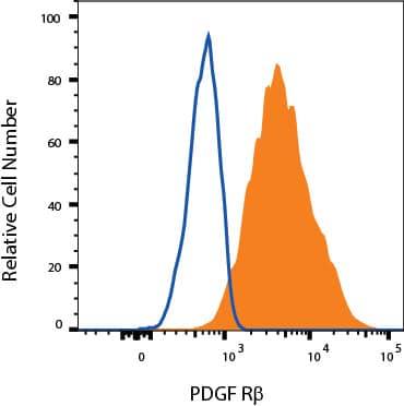
Detection of PDGF R beta in MG‑63 Human Cell Line by Flow Cytometry.
MG-63 human osteosarcoma cell line was stained with Mouse Anti-Human PDGF R beta APC-conjugated Monoclonal Antibody (Catalog # FAB1263A, filled histogram) or isotype control antibody (IC002A, open histogram). View our protocol for Staining Membrane-associated Proteins.Applications for Human PDGF R beta APC-conjugated Antibody
Flow Cytometry
Sample: MG‑63 human osteosarcoma cell line
Formulation, Preparation, and Storage
Purification
Formulation
Shipping
Stability & Storage
- 12 months from date of receipt, 2 to 8 °C as supplied.
Background: PDGF R beta
PDGF is a major serum mitogen that can exist as a homo or hetero-dimeric protein consisting of disulfide-linked PDGF-A and PDGF‑B chains. The PDGF-AA, PDGF‑BB and PDGF-AB isoforms have been shown to bind to two distinct cell surface PDGF receptors with different affinities. Where as PDGF R alpha binds all three PDGF isoforms with high affinity, PDGF R beta binds PDGF-BB only with high-affinity. Both PDGF R alpha and PDGF R beta are members of the class III subfamily of receptor tyrosine kinases (RTK) that also includes the receptors for M-CSF, SCF and Flt3 ligand. All class III RTKs are characterized by the presence of five immunoglobulin-like domains in their extracellular region and a split kinase domain in their intracellular region. PDGF binding induces receptor homo-and hetero‑dimerization and signal transduction. The expression of the alpha and beta receptors is independently regulated in various cell types. Recombinant soluble PDGF R beta binds PDGF with high affinity and is a potent PDGF antagonist (4).
References
- Hart et al. (1987) J. Biol. Chem. 262:10780.
- Gronwald et al. (1988) Proc. Natl. Acad. Sci. 85:3435.
- Seifert et al. (1989) J. Biol. Chem. 264:8771.
- Heldin, C.H. and L. Claesson-Welsh (1994) in Guidebook to Cytokines and Their Receptors, Nicola, N.A. ed. Oxford University Press, New York, p. 202.
Long Name
Alternate Names
Gene Symbol
Additional PDGF R beta Products
Product Documents for Human PDGF R beta APC-conjugated Antibody
Product Specific Notices for Human PDGF R beta APC-conjugated Antibody
For research use only