Human Perforin Antibody
R&D Systems, part of Bio-Techne | Catalog # MAB103853


Conjugate
Catalog #
Key Product Details
Species Reactivity
Human
Applications
Intracellular Staining by Flow Cytometry
Label
Unconjugated
Antibody Source
Monoclonal Mouse IgG2A Clone # 1031751
Product Specifications
Immunogen
Chinese Hamster Ovary cell line CHO-derived human Perforin
Pro22-Trp555
Accession # P14222
Pro22-Trp555
Accession # P14222
Specificity
Detects human Perforin in direct ELISAs.
Clonality
Monoclonal
Host
Mouse
Isotype
IgG2A
Scientific Data Images for Human Perforin Antibody
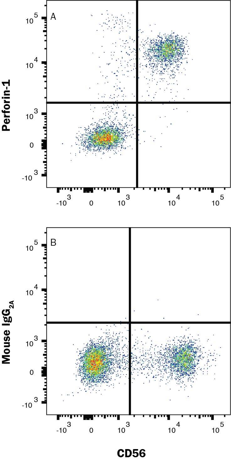
Detection of Perforin in Human PBMCs by Flow Cytometry.
Human peripheral blood mononuclear cells (PBMCs) were stained with (A) Mouse Anti-Human Perforin Monoclonal Antibody (Catalog # MAB103853) or (B) isotype control antibody (MAB002) followed by Anti-Mouse IgG Allophycocyanin-conjugated Secondary Antibody (F0101B) and Mouse anti-Human CD56 Phycoerythrin-conjugated Monoclonal Antibody (FAB24086P). To facilitate intracellular staining, cells were fixed and permeabilized with FlowX FoxP3 Fixation & Permeabilization Buffer Kit (FC012). Staining was performed using our Staining Intracellular Molecules protocol.Applications for Human Perforin Antibody
Application
Recommended Usage
Intracellular Staining by Flow Cytometry
0.25 µg/mL
Sample: Human PBMC fixed and permeabilized with FlowX FoxP3 Fixation & Permeabilization Buffer Kit (Catalog # FC012)
Sample: Human PBMC fixed and permeabilized with FlowX FoxP3 Fixation & Permeabilization Buffer Kit (Catalog # FC012)
Formulation, Preparation, and Storage
Purification
Protein A or G purified from hybridoma culture supernatant
Reconstitution
Reconstitute at 0.5 mg/mL in sterile PBS. For liquid material, refer to CoA for concentration.
Formulation
Lyophilized from a 0.2 μm filtered solution in PBS with Trehalose. *Small pack size (SP) is supplied either lyophilized or as a 0.2 µm filtered solution in PBS.
Shipping
Lyophilized product is shipped at ambient temperature. Liquid small pack size (-SP) is shipped with polar packs. Upon receipt, store immediately at the temperature recommended below.
Stability & Storage
Use a manual defrost freezer and avoid repeated freeze-thaw cycles.
- 12 months from date of receipt, -20 to -70 °C as supplied.
- 1 month, 2 to 8 °C under sterile conditions after reconstitution.
- 6 months, -20 to -70 °C under sterile conditions after reconstitution.
Background: Perforin
Long Name
Perforin 1 (Pore Forming Protein)
Alternate Names
Cytolysin, FLH2, HPLH2, P1, PFP, PRF1
Gene Symbol
PRF1
UniProt
Additional Perforin Products
Product Documents for Human Perforin Antibody
Product Specific Notices for Human Perforin Antibody
For research use only
Loading...
Loading...
Loading...
Loading...
Loading...