Human Phospho-VEGFR2/KDR/Flk-1 (Y1214) Antibody
R&D Systems, part of Bio-Techne | Catalog # AF1766

Key Product Details
Validated by
Species Reactivity
Validated:
Cited:
Applications
Validated:
Cited:
Label
Antibody Source
Product Specifications
Immunogen
Specificity
Clonality
Host
Isotype
Scientific Data Images for Human Phospho-VEGFR2/KDR/Flk-1 (Y1214) Antibody
Detection of Human Phospho-VEGFR2/KDR/Flk‑1 (Y1214) by Western Blot.
Western Blot shows lysates of A431 human epithelial carcinoma cell line untreated (-) or treated (+) with 100 μM pervanadate (PV) for 10 minutes. PVDF membrane was probed with 1 µg/ml of Rabbit Anti-Human Phospho-VEGFR2/KDR/Flk-1 (Y1214) Antigen Affinity-purified Polyclonal Antibody (Catalog # AF1766) followed by HRP-conjugated Anti-Rabbit IgG Secondary Antibody (Catalog # HAF008). A specific band was detected for VEGFR2/KDR/Flk-1 at approximately 230 kDa (as indicated). This experiment was conducted under reducing conditions and using Western Blot Buffer Group 1.Phospho-VEGFR2/KDR/Flk-1 (Y1214) in A431 Human Cell Line.
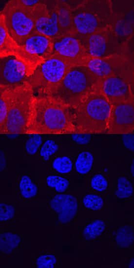
VEGFR2/KDR/Flk-1 phosphorylated at Y1214 was detected in immersion fixed A431 human epithelial carcinoma cell line untreated (lower panel) or treated (upper panel) with pervanadate using Rabbit Anti-Human Phospho-VEGFR2/KDR/Flk-1 (Y1214) Antigen Affinity-purified Polyclonal Antibody (Catalog # AF1766) at 10 µg/mL for 3 hours at room temperature. Cells were stained using the NorthernLights™ 557-conjugated Anti-Rabbit IgG Secondary Antibody (red; NL004) and counterstained with DAPI (blue). View our protocol for Fluorescent ICC Staining of Cells on Coverslips.Detection of Human VEGFR2/KDR/Flk-1 by Western Blot
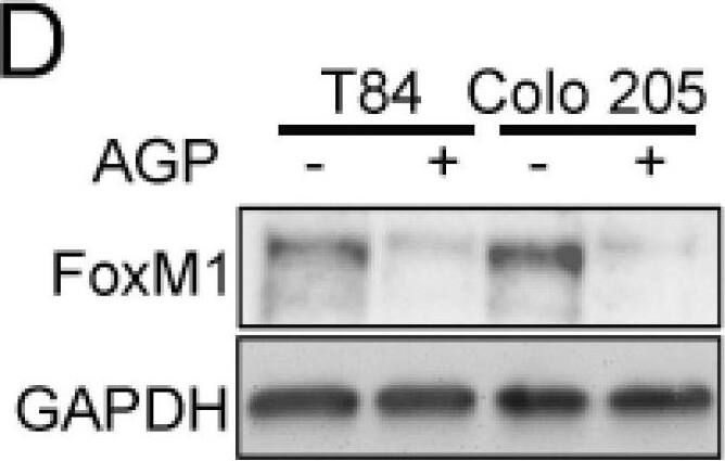
FoxM1 and RASSF1A expression is inversely co-related in colon cancer tissues. (A) Tissue extranction from both normal and colon cancer patient for monitoring the translational level of FoxM1 and RASSF1A by immunoblot. Quantification of RASSF1A (B) and FoxM1 (C) by scanning densitometry. GAPDH was used as a loading control. (D) T84 and Colo 205 cells were treated with or without AGP IC50 (45 µM) for 48 h. Cell lysates were analyzed by Western blot for FoxM1 and GAPDH expression. (E) Quantitative estimations of FoxM1 levels determined by densitometry measurements of western blots from three independent experiments after normalization with GAPDH (p < 0.001). T84 and Colo 205 cells were treated with or without AGP as indicated before and the transcriptional level were determined by qRT-PCR for (F) FoxM1 and (G) PTTG1. Bar graphs show quantitative results normalized to GAPDH mRNA levels. Results are from three independent experiments and statistical significance was determined using one way-ANOVA followed Bonferroni test. (* p < 0.05, ** p < 0.01, *** p < 0.001). Image collected and cropped by CiteAb from the following publication (https://pubmed.ncbi.nlm.nih.gov/30744076), licensed under a CC-BY license. Not internally tested by R&D Systems.Applications for Human Phospho-VEGFR2/KDR/Flk-1 (Y1214) Antibody
Immunocytochemistry
Sample: Immersion fixed A431 human epithelial carcinoma cell line treated with pervanadate
Western Blot
Sample: A431 human epithelial carcinoma cell line untreated (-) or treated (+) with 100 μM pervanadate (PV) for 10 minutes
Formulation, Preparation, and Storage
Purification
Reconstitution
Formulation
*Small pack size (-SP) is supplied either lyophilized or as a 0.2 µm filtered solution in PBS.
Shipping
Stability & Storage
- 12 months from date of receipt, -20 to -70 °C as supplied.
- 1 month, 2 to 8 °C under sterile conditions after reconstitution.
- 6 months, -20 to -70 °C under sterile conditions after reconstitution.
Background: VEGFR2/KDR/Flk-1
VEGFR2 (KDR/Flk-1), VEGFR1 (Flt-1) and VEGFR3 (Flt-4) belong to the class III subfamily of receptor tyrosine kinases (RTKs). All three receptors contain seven immunoglobulin-like repeats in their extracellular domains and kinase insert domains in their intracellular regions. The expression of VEGFR1, 2, and 3 is almost exclusively restricted to the endothelial cells. These receptors are likely to play essential roles in vasculogenesis and angiogenesis.
VEGFR2 cDNA encodes a 1356 amino acid (aa) residue precursor protein with a 19 aa residue signal peptide. Mature VEGFR2 is composed of a 745 aa residue extracellular domain, a 25 aa residue transmembrane domain and a 567 aa residue cytoplasmic domain. In contrast to VEGFR1 which binds both PlGF and VEGF with high affinity, VEGFR2 binds VEGF but not PlGF with high affinity. The recombinant soluble VEGFR2/Fc chimera binds VEGF with high affinity and is a potent VEGF antagonist.
References
- Ferra, N. and R. Davis-Smyth (1997) Endocrine Reviews 18:4.
Long Name
Alternate Names
Gene Symbol
Additional VEGFR2/KDR/Flk-1 Products
Product Documents for Human Phospho-VEGFR2/KDR/Flk-1 (Y1214) Antibody
Product Specific Notices for Human Phospho-VEGFR2/KDR/Flk-1 (Y1214) Antibody
For research use only


