Human PILR-alpha Antibody
R&D Systems, part of Bio-Techne | Catalog # MAB64841


Key Product Details
Species Reactivity
Applications
Label
Antibody Source
Product Specifications
Immunogen
Gln20-Thr196
Accession # Q9UKJ1
Specificity
Clonality
Host
Isotype
Scientific Data Images for Human PILR-alpha Antibody
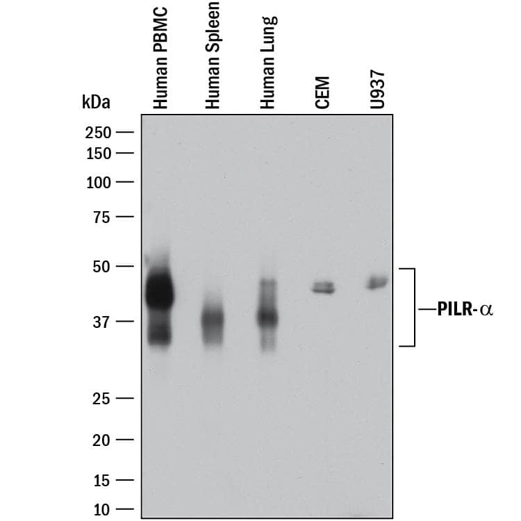
Detection of Human PILR‑ alpha by Western Blot.
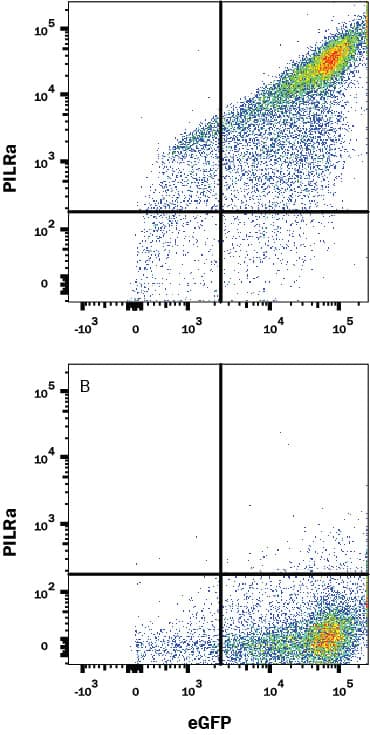
Western blot shows lysates of human peripheral blood mononuclear cells (PBMCs), human spleen tissue, human lung tissue, CEM human T-lymphoblastoid cell line, and U937 human histiocytic lymphoma cell line. PVDF membrane was probed with 2 µg/mL of Rabbit Anti-Human PILR-a Monoclonal Antibody (Catalog # MAB64841) followed by HRP-conjugated Anti-Rabbit IgG Secondary Antibody (Catalog # HAF008). A specific band was detected for PILR-a at approximately 35-50 kDa (as indicated). This experiment was conducted under reducing conditions and using Immunoblot Buffer Group 1.Detection of PILR-alpha R in HEK293 Human Cell Line Transfected with Human PILR-alpha and eGFP by Flow Cytometry
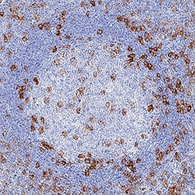
HEK293 human embryonic kidney cell line transfected with either (A) human PILR-alpha or (B) irrelevant transfectants and eGFP was stained with Rabbit Anti-Human PILR-alpha Monoclonal Antibody (Catalog # MAB64841) followed by Allophycocyanin-conjugated Anti-Rabbit IgG Secondary Antibody (Catalog # F0111). Quadrant markers were set based on control antibody staining (Catalog # MAB1050). View our protocol for Staining Intracellular Molecules.PILR‑ alpha in Human Tonsil.
PILR-a was detected in immersion fixed paraffin-embedded sections of human tonsil using Rabbit Anti-Human PILR-a Monoclonal Antibody (Catalog # MAB64841) at 3 µg/mL for 1 hour at room temperature followed by incubation with the Anti-Rabbit IgG VisUCyte™ HRP Polymer Antibody (Catalog # VC003). Tissue was stained using DAB (brown) and counterstained with hematoxylin (blue). Specific staining was localized to dendritic cells. View our protocol for IHC Staining with VisUCyte HRP Polymer Detection Reagents.Applications for Human PILR-alpha Antibody
Flow Cytometry
Sample: HEK293 Human Cell Line Transfected with Human PILR-alpha and eGFP
Immunohistochemistry
Sample: Immersion fixed paraffin-embedded sections of human tonsil
Western Blot
Sample: Human peripheral blood mononuclear cells (PBMCs), human spleen tissue, human lung tissue, CEM human T-lymphoblastoid cell line, and U937 human histiocytic lymphoma cell line
Reviewed Applications
Read 1 review rated 5 using MAB64841 in the following applications:
Formulation, Preparation, and Storage
Purification
Reconstitution
Formulation
Shipping
Stability & Storage
- 12 months from date of receipt, -20 to -70 °C as supplied.
- 1 month, 2 to 8 °C under sterile conditions after reconstitution.
- 6 months, -20 to -70 °C under sterile conditions after reconstitution.
Background: PILR-alpha
Paired immunoglobulin-like type 2 receptor alpha (PILRa; also inhibitory receptor PILR-alpha) are 44-50 kDa paired receptors that consist of highly related activating and inhibitory receptors, and are widely involved in the regulation of the immune system. PILR-alpha is thought to act as a cellular signaling inhibitory receptor by recruiting cytoplasmic phosphatases like PTPN6/SHP-1 and PTPN11/SHP-2 via their SH2 domains that block signal transduction through dephosphorylation of signaling molecules. Human PILR-alpha is synthesized as a 303 amino acid (aa) precursor that contains a 19 aa signal sequence, a 178 aa extracellular domain (ECD), a 21 aa transmembrane segment, and an 85 aa cytoplasmic domain. The ECD contains one Ig-like V-type domain and one potential site for N-linked glycosylation. The cytoplasmic domain contains two ITIM motifs (aa 267-272 and 296-301). Alternate splicing generates multiple shorter isoforms. One is TM and possesses a 35 aa substitution for aa 264-303, while others are soluble, and show a deletion of aa 152-224 that may be coupled to the 35 aa substitution noted above, or simply exhibit a 24 aa substitution for aa 152-303. Mature human PILR-alpha is 45% aa identical to mature mouse PILR-alpha. PILR-alpha is predominantly detected in hemopoietic tissues and is expressed in monocytes, macrophages, and granulocytes, but not lymphocytes. It is also strongly expressed by dendritic cells. PILR-alpha interacts with herpes simplex 1 glycoprotein B and functions as an entry coreceptor for this virus.
Long Name
Alternate Names
Gene Symbol
UniProt
Additional PILR-alpha Products
Product Documents for Human PILR-alpha Antibody
Product Specific Notices for Human PILR-alpha Antibody
For research use only

