Human PLZF Antibody
R&D Systems, part of Bio-Techne | Catalog # AF2944


Key Product Details
Species Reactivity
Validated:
Human
Cited:
Human, Mouse, Rat, Transgenic Mouse
Applications
Validated:
Simple Western, Western Blot
Cited:
Flow Cytometry, IHC-F, Immunocytochemistry, Immunohistochemistry, Immunohistochemistry-Paraffin, Immunoprecipitation, Western Blot
Label
Unconjugated
Antibody Source
Polyclonal Goat IgG
Product Specifications
Immunogen
E. coli-derived recombinant human PLZF
Met1-Gln254
Accession # Q05516
Met1-Gln254
Accession # Q05516
Specificity
Detects human PLZF in direct ELISAs and Western blots.
Clonality
Polyclonal
Host
Goat
Isotype
IgG
Scientific Data Images for Human PLZF Antibody
Detection of Human PLZF by Western Blot.
Western blot shows lysates of HUVEC human umbilical vein endothelial cells, HEL 92.1.7 human erythroleukemic cell line, LNCaP human prostate cancer cell line, and 293T human embryonic kidney cell line. PVDF membrane was probed with 1 µg/mL of Goat Anti-Human PLZF Antigen Affinity-purified Polyclonal Antibody (Catalog # AF2944) followed by HRP-conjugated Anti-Goat IgG Secondary Antibody (Catalog # HAF019). A specific band was detected for PLZF at approximately 85 kDa (as indicated). This experiment was conducted under reducing conditions and using Immunoblot Buffer Group 1.Detection of Human PLZF by Simple WesternTM.
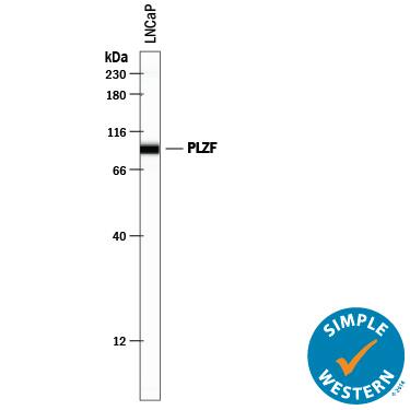
Simple Western lane view shows lysates of LNCaP human prostate cancer cell line, loaded at 0.2 mg/mL. A specific band was detected for PLZF at approximately 94 kDa (as indicated) using 10 µg/mL of Goat Anti-Human PLZF Antigen Affinity-purified Polyclonal Antibody (Catalog # AF2944) followed by 1:50 dilution of HRP-conjugated Anti-Goat IgG Secondary Antibody (Catalog # HAF109). This experiment was conducted under reducing conditions and using the 12-230 kDa separation system.Detection of Mouse PLZF by Western Blot
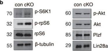
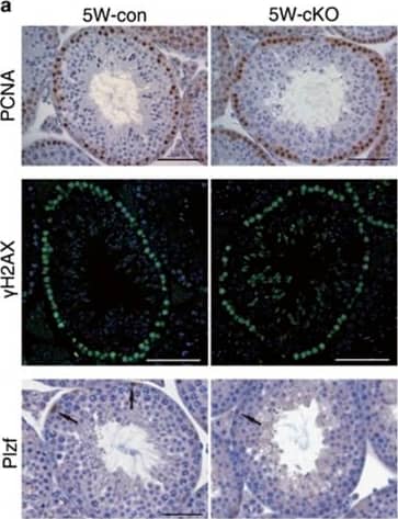
Increased mTORC1 activity in germ cells from Lkb1 newborn cKO testis. (a) Western blot of testis proteins at different postnatal ages using specific antibodies against p-S6K1 (T389), p-rpS6 (S235/6), rpS6 and beta-tubulin. rpS6, and beta-tubulin were used as internal controls. (b) Expression of PI3K-, mTOR- and SPC-related markers by western blot in P8 testes from control and Lkb1 cKO mice. (c) Immunohistochemistry for VASA, GATA4, p-rpS6 in P8 control testis. (d) Immunohistochemistry for Lin28a and p-rpS6 in P8 control testis. Serial sections were used in order to compare cellular localizations of these markers in the same tubule. Asterisks, different distribution of Lin28a and p-rpS6 in the same tubule resulting in strong positive for Lin28a and weak staining for p-rpS6. Arrows, cells expressing both Lin28a and p-rpS6. (e) Immunohistochemistry for Plzf and p-rpS6 in control and cKO mice. (f) Mean number of Plzf- or p-rpS6-positive cells in each tubule (positive cells/total tubules). The data were shown as mean±S.E.M. of at least three replicates. *P<0.05. Scale bars=50 μm Image collected and cropped by CiteAb from the following publication (https://pubmed.ncbi.nlm.nih.gov/29022902), licensed under a CC-BY license. Not internally tested by R&D Systems.Applications for Human PLZF Antibody
Application
Recommended Usage
Simple Western
10 µg/mL
Sample: LNCaP human prostate cancer cell line
Sample: LNCaP human prostate cancer cell line
Western Blot
1 µg/mL
Sample: HUVEC human umbilical vein endothelial cells, HEL 92.1.7 human erythroleukemic cell line, LNCaP human prostate cancer cell line, and 293T human embryonic kidney cell line
Sample: HUVEC human umbilical vein endothelial cells, HEL 92.1.7 human erythroleukemic cell line, LNCaP human prostate cancer cell line, and 293T human embryonic kidney cell line
Formulation, Preparation, and Storage
Purification
Antigen Affinity-purified
Reconstitution
Reconstitute at 0.2 mg/mL in sterile PBS. For liquid material, refer to CoA for concentration.
Formulation
Lyophilized from a 0.2 μm filtered solution in PBS with Trehalose. *Small pack size (SP) is supplied either lyophilized or as a 0.2 µm filtered solution in PBS.
Shipping
Lyophilized product is shipped at ambient temperature. Liquid small pack size (-SP) is shipped with polar packs. Upon receipt, store immediately at the temperature recommended below.
Stability & Storage
Use a manual defrost freezer and avoid repeated freeze-thaw cycles.
- 12 months from date of receipt, -20 to -70 °C as supplied.
- 1 month, 2 to 8 °C under sterile conditions after reconstitution.
- 6 months, -20 to -70 °C under sterile conditions after reconstitution.
Background: PLZF
Long Name
Promyelocytic Leukemia Zinc Finger
Alternate Names
ZBTB16, ZNF145
Gene Symbol
ZBTB16
UniProt
Additional PLZF Products
Product Documents for Human PLZF Antibody
Product Specific Notices for Human PLZF Antibody
For research use only
Loading...
Loading...
Loading...
Loading...


