Human PSGL-1/CD162 PE-conjugated Antibody
R&D Systems, part of Bio-Techne | Catalog # FAB9961P


Key Product Details
Species Reactivity
Applications
Label
Antibody Source
Product Specifications
Immunogen
Gln42-Gly295
Accession # NP_002997
Specificity
Clonality
Host
Isotype
Scientific Data Images for Human PSGL-1/CD162 PE-conjugated Antibody
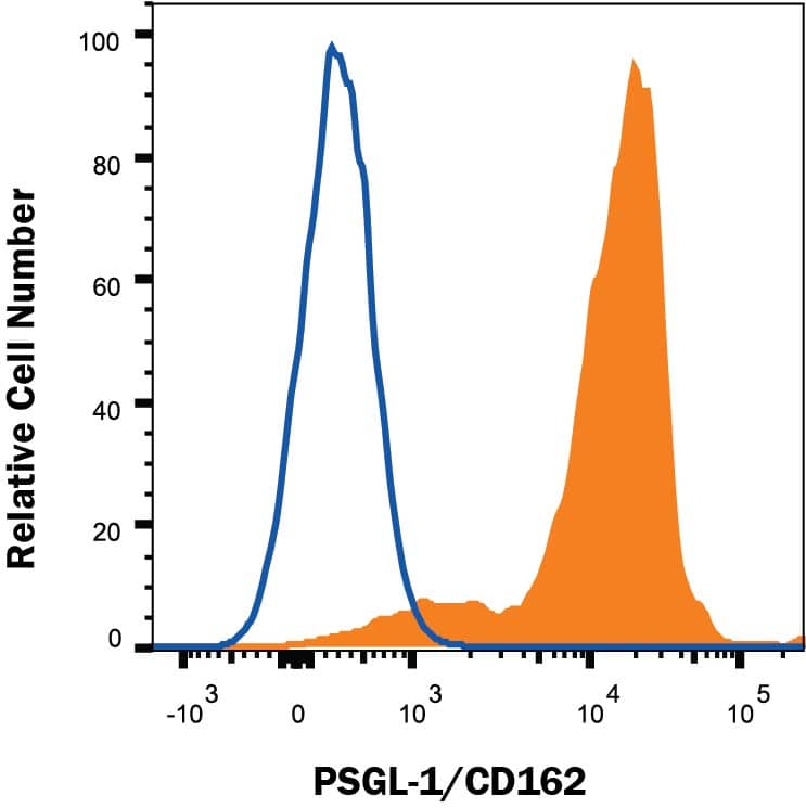
Detection of PSGL‑1/CD162 in Human Peripheral Blood Lymphocytes by Flow Cytometry.
Human peripheral blood lymphocytes were stained with Mouse Anti-Human PSGL-1/CD162 PE-conjugated Monoclonal Antibody (Catalog # FAB9961P, filled histogram) or isotype control antibody (Catalog # IC003P, open histogram). View our protocol for Staining Membrane-associated Proteins.Applications for Human PSGL-1/CD162 PE-conjugated Antibody
Flow Cytometry
Sample: Human peripheral blood lymphocytes
Formulation, Preparation, and Storage
Purification
Formulation
Shipping
Stability & Storage
Background: PSGL-1/CD162
Human PSGL-1 (P-Selectin Glycoprotein Ligand-1; also CD162), is a 120 kDa mucin-type glycoprotein that plays a key role in leukocyte adhesion (1-3). It is synthesized as a 412 amino acid (aa) preproprecursor that contains a 17 aa signal sequence, a 24 aa propeptide, a 279 aa extracellular domain (ECD), a 21 aa transmembrane segment and a 71 aa cytoplasmic region (4, 5). Following cleavage of the pre- and prosegments, it is expressed as a 240 kDa disulfide-linked homodimer. The extreme N-terminus (aa 1-16 of the mature molecule) contains one threonine (aa 16) and three tyrosines (aa 5, 7, and 10) that are involved in ligand binding. The Thr residue allows for O-linked glycosylation in the form of a core-2 structure (GalNAc-Gal) linked in a beta1,6 bond to a sialylated Lewis X motif (GlcNAc linked to both Fuc and Gal with a terminal sialic acid residue) (1, 2, 5, 6, 7). The three tyrosine residues allow for sulfation (8, 9). When binding to P-selectin, Tyr sulfation and glycosylation are essential. Tyr7 provides the most efficient sulfate moiety, while Fuc and sialic acid are essentially mandatory (7). When binding to E-selectin, only carbohydrate is needed, while both carbohydrate and Tyr10 are used for L-selectin binding (6, 8). There are 16 decameric aa repeats in the ECD of the longform of PSGL-1. This form is referred to as the A allele, and represents 65 - 80% of the population. Alleles B and C show deletions of decameric repeats #2 (aa 132-141) plus #9 and 10 (aa 222-241), respectively. Shorter forms may show weaker binding to P-selectin (9, 10). Soluble forms of PSGL-1 are also known. Neutrophil elastase will cleave somewhere within repeats #5-9, while cathepsin G cleaves after Tyr7 (11). The loss of Tyr5 and 7 should impact binding affinity. PSGL-1 is found on virtually all leukocytes and macrophages/DC’s (1). Although there is similarity in the organization of the ECD between species, there is little aa identity. Human PSGL-1 ECD shares 51%, 52% and 43% aa sequence identity with equine, canine and mouse ECD, respectively.
References
- Yang, J. et al. (1999) Thromb. Haemost. 81:1.
- Cummings, R.D. (1999) Braz. J. Med. Biol. Res. 32:519.
- McEver, R.P. and R.D. Cummings (1997) J. Clin. Invest. 100:485.
- Sako, D. et al. (1993) Cell 75:1179.
- Veldman, G.M. et al. (1995) J. Biol. Chem. 270:16470.
- Bernimoulin, M.P. et al. (2003) J. Biol. Chem. 278:37.
- Leppanen, A. et al. (2000) J. Biol. Chem. 275:39569.
- Sako, D. et al. (1995) Cell 83:323.
- Afshar-Kharghan, V. et al. (2001) Blood 97:3306.
- Lozano, M.L. et al. (2001) Br. J. Haematol. 115:969.
- Gardiner, E.E. et al. (2001) Blood 98:1440.
Long Name
Alternate Names
Gene Symbol
UniProt
Additional PSGL-1/CD162 Products
Product Documents for Human PSGL-1/CD162 PE-conjugated Antibody
Product Specific Notices for Human PSGL-1/CD162 PE-conjugated Antibody
For research use only