Human PTP1B Antibody
R&D Systems, part of Bio-Techne | Catalog # AF1366

Key Product Details
Species Reactivity
Validated:
Cited:
Applications
Validated:
Cited:
Label
Antibody Source
Product Specifications
Immunogen
Specificity
Clonality
Host
Isotype
Scientific Data Images for Human PTP1B Antibody
PTP1B in Human Breast Cancer Tissue.
PTP1B was detected in immersion fixed paraffin-embedded sections of human breast cancer tissue using Rabbit Anti-Human PTP1B Antigen Affinity-purified Polyclonal Antibody (Catalog # AF1366) at 15 µg/mL overnight at 4 °C. Tissue was stained using the Anti-Rabbit HRP-DAB Cell & Tissue Staining Kit (brown; Catalog # CTS005) and counterstained with hematoxylin (blue). Specific labeling was localized to the perinuclear region in glandular epithelial cells. View our protocol for Chromogenic IHC Staining of Paraffin-embedded Tissue Sections.Detection of Human PTP1B by Western Blot.
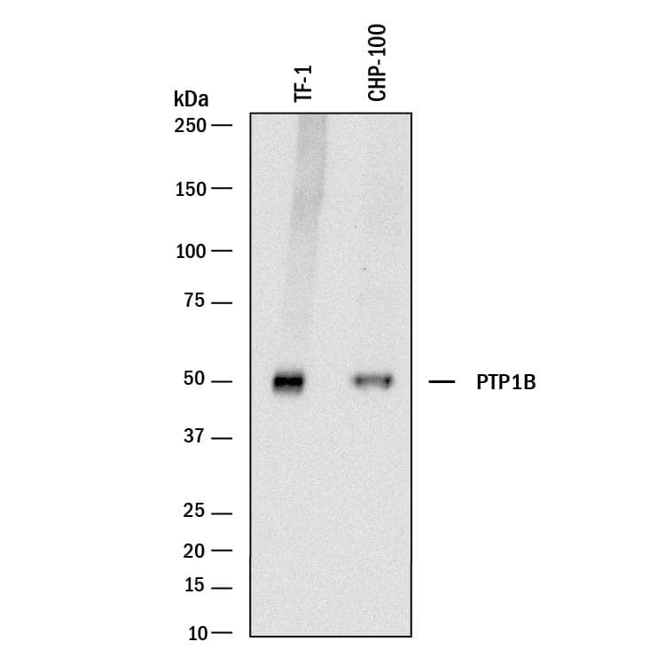
Western blot shows lysates of TF-1 human erythroleukemic cell line and CHP-100 human neuroblastoma cell line. PVDF membrane was probed with 0.5 µg/mL of Rabbit Anti-Human PTP1B Antigen Affinity-purified Polyclonal Antibody (Catalog # AF1366) followed by HRP-conjugated Anti-Rabbit IgG Secondary Antibody (Catalog # HAF008). A specific band was detected for PTP1B at approximately 50 kDa (as indicated). This experiment was conducted under reducing conditions and using Immunoblot Buffer Group 1.Detection of Human PTP1B by Simple WesternTM.
Simple Western lane view shows lysates of HeLa human cervical epithelial carcinoma cell line, loaded at 0.2 mg/mL. A specific band was detected for PTP1B at approximately 57 kDa (as indicated) using 10 µg/mL of Rabbit Anti-Human PTP1B Antigen Affinity-purified Polyclonal Antibody (Catalog # AF1366). This experiment was conducted under reducing conditions and using the 12-230 kDa separation system.Applications for Human PTP1B Antibody
Immunohistochemistry
Sample: Immersion fixed paraffin-embedded sections of human breast cancer tissue
Simple Western
Sample: HeLa human cervical epithelial carcinoma cell line
Western Blot
Sample: TF‑1 human erythroleukemic cell line and CHP‑100 human neuroblastoma cell line
Reviewed Applications
Read 1 review rated 4 using AF1366 in the following applications:
Formulation, Preparation, and Storage
Purification
Reconstitution
Formulation
Shipping
Stability & Storage
- 12 months from date of receipt, -20 to -70 °C as supplied.
- 1 month, 2 to 8 °C under sterile conditions after reconstitution.
- 6 months, -20 to -70 °C under sterile conditions after reconstitution.
Background: PTP1B
Protein tyrosine phosphatase 1B (PTP1B) is an enzyme that removes phosphate groups covalently attached to tyrosine residues in proteins. This ubiquitously expressed enzyme is anchored in the endoplasmic reticulum by its C-terminal end and has its catalytic regions exposed to the cytosol. The recombinant protein lacks the C-terminal 114 amino acids but is fully active. PTP1B will dephosphorylate a wide variety of phosphoproteins, such as receptors for the growth factors insulin and epidermal growth factor (EGF), c-Src and beta-catenin. Of particular interest is the observation that PTP1B knock-out mice are resistant to high-caloric intake-induced obesity and have exaggerated insulin responses, suggesting that PTP1B may play an important role in regulating growth factor responsiveness.
References
- Angers-Loustau, et al. (1999) Biochem. Cell Biol. 77:493.
- Sarmiento, et al. (1998) J. Biol. Chem. 273:26368.
- Bjorge, et al. (2000) J. Biol. Chem. 52:41439.
- Balsamo, et al. (1996) J. Cell Biol. 134:801.
- Elchebly, et al. (1999) Science 283:1544.
Long Name
Alternate Names
Gene Symbol
Additional PTP1B Products
Product Documents for Human PTP1B Antibody
Product Specific Notices for Human PTP1B Antibody
For research use only


