Human RAMP1 Antibody
R&D Systems, part of Bio-Techne | Catalog # AF6428

Key Product Details
Species Reactivity
Applications
Label
Antibody Source
Product Specifications
Immunogen
Cys27-Ser117
Accession # O60894
Specificity
Clonality
Host
Isotype
Scientific Data Images for Human RAMP1 Antibody
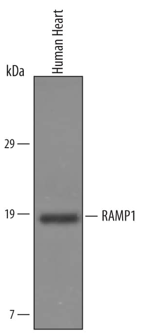
Detection of Human RAMP1 by Western Blot.
Western blot shows lysates of human heart tissue. PVDF Membrane was probed with 1 µg/mL of Sheep Anti-Human RAMP1 Antigen Affinity-purified Polyclonal Antibody (Catalog # AF6428) followed by HRP-conjugated Anti-Sheep IgG Secondary Antibody (Catalog # HAF016). A specific band was detected for RAMP1 at approximately 17 kDa (as indicated). This experiment was conducted under reducing conditions and using Immunoblot Buffer Group 1.RAMP1 in Human Brain.
RAMP1 was detected in immersion fixed paraffin-embedded sections of human brain (hypothalamus) using Sheep Anti-Human RAMP1 Antigen Affinity-purified Polyclonal Antibody (Catalog # AF6428) at 10 µg/mL overnight at 4 °C. Before incubation with the primary antibody, tissue was subjected to heat-induced epitope retrieval using Antigen Retrieval Reagent-Basic (Catalog # CTS013). Tissue was stained using the Anti-Sheep HRP-DAB Cell & Tissue Staining Kit (brown; Catalog # CTS019) and counterstained with hematoxylin (blue). Specific staining was localized to neuronal and glial processes. View our protocol for Chromogenic IHC Staining of Paraffin-embedded Tissue Sections.RAMP1 in Human Thyroid Cancer Tissue..
RAMP1 was detected in immersion fixed paraffin-embedded sections of human thyroid cancer tissue using Sheep Anti-Human RAMP1 Antigen Affinity-purified Polyclonal Antibody (Catalog # AF6428) at 15 µg/mL for 1 hour at room temperature followed by incubation with the Anti-Sheep IgG VisUCyte™ HRP Polymer Antibody (VC006). Before incubation with the primary antibody, tissue was subjected to heat-induced epitope retrieval using Antigen Retrieval Reagent-Basic (CTS013). Tissue was stained using DAB (brown) and counterstained with hematoxylin (blue). Specific staining was localized to vasculature. Staining was performed using our protocol forIHC Staining with VisUCyte HRP Polymer Detection Reagents.Applications for Human RAMP1 Antibody
Immunohistochemistry
Sample: Immersion fixed paraffin-embedded sections of human brain (hypothalamus) and immersion fixed paraffin-embedded sections of human thyroid cancer tissue
Western Blot
Sample: Human heart tissue
Formulation, Preparation, and Storage
Purification
Reconstitution
Formulation
Shipping
Stability & Storage
- 12 months from date of receipt, -20 to -70 °C as supplied.
- 1 month, 2 to 8 °C under sterile conditions after reconstitution.
- 6 months, -20 to -70 °C under sterile conditions after reconstitution.
Background: RAMP1
RAMP1 (Receptor Activity Modifying Protein 1) is a 14‑18 kDa member of the RAMP family of proteins. Variability in MW is due to the absence, or presence, of intramolecular disulfide bonds that form in the ER and Golgi. It is expressed on/in neurons, vascular endothelial cells, visceral and vascular smooth muscle cells, mammary epithelium, osteoblasts and cardiomyocytes. RAMP1 interacts with CRLR/CLR to form an 84 kDa noncovalent receptor complex for CGRP and AM, and with CTR to form a 76 kDa receptor complex for amylin. Mature human RAMP1 is a 122 amino acid (aa) nonglycosylated type I transmembrane protein that contains a 91 aa extracellular domain (ECD) (aa 27‑117) plus a ten aa cytoplasmic tail. In the ECD, residues 78‑90 bind AM, while residues 91‑103 bind CGRP. In the absence of CRLR and CTR, RAMP1 will form 30‑32 kDa disulfide‑linked homodimers in the ER/Golgi. Over aa 27‑117, human RAMP1 shares 65% aa identity with mouse RAMP1.
Long Name
Alternate Names
Gene Symbol
UniProt
Additional RAMP1 Products
Product Documents for Human RAMP1 Antibody
Product Specific Notices for Human RAMP1 Antibody
For research use only


