Human/Rat/Hamster ACE-2 Antibody
R&D Systems, part of Bio-Techne | Catalog # MAB9331

Key Product Details
Species Reactivity
Validated:
Cited:
Applications
Validated:
Cited:
Label
Antibody Source
Product Specifications
Immunogen
Gln18-Ser740 (predicted)
Accession # Q9BYF1
Specificity
Clonality
Host
Isotype
Scientific Data Images for Human/Rat/Hamster ACE-2 Antibody
Detection of Human ACE‑2 by Western Blot.
Western blot shows lysates of NS0 mouse myeloma cell line and human kidney tissue. PVDF membrane was probed with 2 µg/mL of Mouse Anti-Human ACE-2 Monoclonal Antibody (Catalog # MAB9331) followed by HRP-conjugated Anti-Mouse IgG Secondary Antibody (Catalog # HAF007). A specific band was detected for ACE-2 at approximately 110 kDa (as indicated). This experiment was conducted under reducing conditions and using Immunoblot Buffer Group 1.ACE-2 in Rat Kidney.
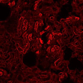
ACE-2 was detected in immersion fixed frozen sections of rat kidney using Mouse Anti-Human ACE-2 Monoclonal Antibody (Catalog # MAB9331) at 1 µg/mL overnight at 4 °C. Before incubation with the primary antibody, tissue was subjected to heat-induced epitope retrieval using Antigen Retrieval Reagent-Basic (CTS013). Tissue was stained using the NorthernLights™ 557-conjugated Anti-Mouse IgG Secondary Antibody (red; NL007). Specific staining was localized to convoluted tubules. Staining was performed using our protocol for Fluorescent IHC Staining of Frozen Tissue Sections.ACE-2 in Hamster Lung.
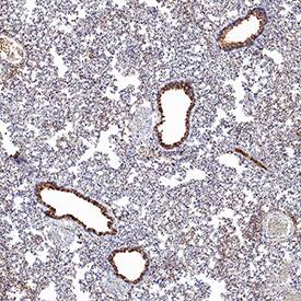
ACE-2 was detected in immersion fixed paraffin-embedded sections of hamster lung using Mouse Anti-Human ACE-2 Monoclonal Antibody (Catalog # MAB9331) at 10 µg/mL for 1 hour at room temperature followed by incubation with the Anti-Mouse IgG VisUCyte™ HRP Polymer Antibody (VC001). Before incubation with the primary antibody, tissue was subjected to heat-induced epitope retrieval using Antigen Retrieval Reagent-Basic (CTS013). Tissue was stained using DAB (brown) and counterstained with hematoxylin (blue). Specific staining was localized to respiratory bronchioles. Staining was performed using our protocol for IHC Staining with VisUCyte HRP Polymer Detection Reagents.Applications for Human/Rat/Hamster ACE-2 Antibody
Immunohistochemistry
Sample: Frozen sections of rat kidney and paraffin-embedded sections of hamster lung.
Immunoprecipitation
Sample: Conditioned cell culture medium spiked with Recombinant Human ACE-2 (Catalog # 933-ZN), see our available Western blot detection antibodies
Western Blot
Sample: NS0 mouse myeloma cell line and human kidney tissue
Reviewed Applications
Read 1 review rated 5 using MAB9331 in the following applications:
Formulation, Preparation, and Storage
Purification
Reconstitution
Formulation
Shipping
Stability & Storage
- 12 months from date of receipt, -20 to -70 °C as supplied.
- 1 month, 2 to 8 °C under sterile conditions after reconstitution.
- 6 months, -20 to -70 °C under sterile conditions after reconstitution.
Background: ACE-2
Angiogensin I Converting Enzyme-2 (ACE-2) is a type I transmembrane zinc protease that cleaves angiotensins I and II to produce vasodilatory and anti-proliferative peptides. The balance between ACE-1 and ACE-2 activity is critical for maintaining cardiovascular, renal, and pulmonary function. ACE-2 isoforms of 75 kDa and 120 kDa are differentially expressed between lung and kidney, respectively, and a shed soluble form is generated by TACE/ADAM17 mediated cleavage. ACE-2 also functions as the cellular uptake receptor for the SARS coronoavirus. Within the extracellular domain, human ACE-2 shares 83% aa sequence identity with mouse and rat ACE-2.
Long Name
Alternate Names
Entrez Gene IDs
Gene Symbol
UniProt
Additional ACE-2 Products
Product Documents for Human/Rat/Hamster ACE-2 Antibody
Product Specific Notices for Human/Rat/Hamster ACE-2 Antibody
For research use only


