Human Semaphorin 7A Antibody
R&D Systems, part of Bio-Techne | Catalog # AF2068


Key Product Details
Species Reactivity
Validated:
Human
Cited:
Human
Applications
Validated:
CyTOF-ready, Flow Cytometry, Western Blot
Cited:
Immunocytochemistry, Immunohistochemistry, Immunoprecipitation, Western Blot
Label
Unconjugated
Antibody Source
Polyclonal Goat IgG
Product Specifications
Immunogen
Mouse myeloma cell line NS0-derived recombinant human Semaphorin 7A
His47-Ala648
Accession # O75326
His47-Ala648
Accession # O75326
Specificity
Detects human Semaphorin 7A in direct ELISAs and Western blots. In direct ELISAs, approximately 50% cross-reactivity with recombinant mouse Semaphorin 7A is observed.
Clonality
Polyclonal
Host
Goat
Isotype
IgG
Scientific Data Images for Human Semaphorin 7A Antibody
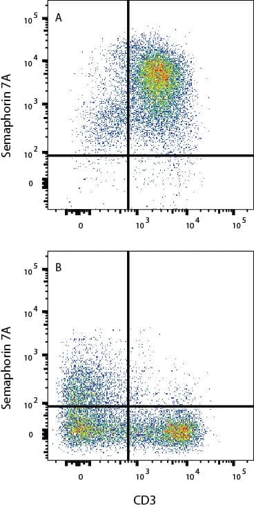
Detection of Semaphorin 7A in PBMC lymphocytes treated +/- 5 µg/mL PHA for 2 days by Flow Cytometry
PBMC lymphocytes treated with 5 µg/mL PHA for 2 days (A) vs naïve PBMC lymphocytes (B) were stained with Goat Anti-Human Semaphorin 7A Antigen Affinity-purified Polyclonal Antibody (Catalog # AF2068) followed by Phycoerythrin-conjugated Anti-Goat IgG Secondary Antibody (Catalog # F0107) and Mouse anti-Human CD3 APC-conjugated Monoclonal antibody (Catalog # FAB100A). View our protocol for Staining Membrane-associated Proteins.Detection of Semaphorin 7A in HT1080 cells by Flow Cytometry
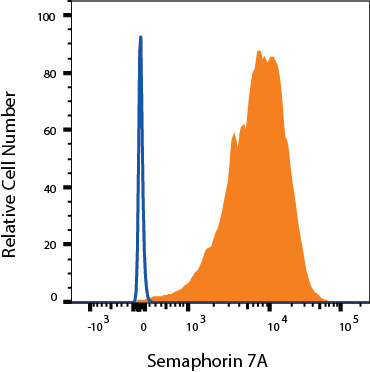
HT1080 cells were stained with Goat Anti-Human Semaphorin 7A Antigen Affinity-purified Polyclonal Antibody (Catalog # AF2068, filled histogram) or isotype control antibody (Catalog # AB-108-C, open histogram) followed by Phycoerythrin-conjugated Anti-Goat IgG Secondary Antibody (Catalog # F0107). View our protocol for Staining Membrane-associated Proteins.Applications for Human Semaphorin 7A Antibody
Application
Recommended Usage
CyTOF-ready
Ready to be labeled using established conjugation methods. No BSA or other carrier proteins that could interfere with conjugation.
Flow Cytometry
0.25 µg/106 cells
Sample: Human T cells treated with PHA (see details below); HT1080 human fibrosarcoma cell line
Sample: Human T cells treated with PHA (see details below); HT1080 human fibrosarcoma cell line
Western Blot
0.1 µg/mL
Sample: Recombinant Human Semaphorin 7A
Sample: Recombinant Human Semaphorin 7A
Formulation, Preparation, and Storage
Purification
Antigen Affinity-purified
Reconstitution
Reconstitute at 0.2 mg/mL in sterile PBS. For liquid material, refer to CoA for concentration.
Formulation
Lyophilized from a 0.2 μm filtered solution in PBS with Trehalose. *Small pack size (SP) is supplied either lyophilized or as a 0.2 µm filtered solution in PBS.
Shipping
Lyophilized product is shipped at ambient temperature. Liquid small pack size (-SP) is shipped with polar packs. Upon receipt, store immediately at the temperature recommended below.
Stability & Storage
Use a manual defrost freezer and avoid repeated freeze-thaw cycles.
- 12 months from date of receipt, -20 to -70 °C as supplied.
- 1 month, 2 to 8 °C under sterile conditions after reconstitution.
- 6 months, -20 to -70 °C under sterile conditions after reconstitution.
Background: Semaphorin 7A
Semaphorin 7A (Sema 7A), also known as Sema L, Sema K1 and CD108, is an 80 kDa glycosylphosphatidylinositol-anchored protein belonging to the Semaphorin superfamily. It binds to the beta1 integrin subunit as well as Plexin C1. The Sema domain of human Sema 7A shares 92% and 90% amino acid sequence homology with that of the mouse and canine protein.
Alternate Names
CD108, H-Sema-L, Sema7A, SEMAL
Gene Symbol
SEMA7A
UniProt
Additional Semaphorin 7A Products
Product Documents for Human Semaphorin 7A Antibody
Product Specific Notices for Human Semaphorin 7A Antibody
For research use only
Loading...
Loading...
Loading...
Loading...
Loading...
