Human TIMP-1 Antibody
R&D Systems, part of Bio-Techne | Catalog # AF970


Key Product Details
Validated by
Species Reactivity
Validated:
Cited:
Applications
Validated:
Cited:
Label
Antibody Source
Product Specifications
Immunogen
Cys24-Ala207
Accession # Q6FGX5
Specificity
Clonality
Host
Isotype
Endotoxin Level
Scientific Data Images for Human TIMP-1 Antibody
Detection of Human TIMP‑1 by Western Blot.
Western blot shows lysates of human lung tissue and human prostate tissue. PVDF membrane was probed with 1 µg/mL of Goat Anti-Human TIMP-1 Antigen Affinity-purified Polyclonal Antibody (Catalog # AF970) followed by HRP-conjugated Anti-Goat IgG Secondary Antibody (Catalog # HAF017). A specific band was detected for TIMP-1 at approximately 25 kDa (as indicated). This experiment was conducted under reducing conditions and using Immunoblot Buffer Group 1.Detection of Human TIMP‑1 by Western Blot.
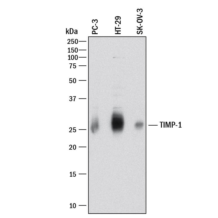
Western blot shows lysates of PC-3 human prostate cancer cell line, HT-29 human colon adenocarcinoma cell line, and SK-OV-3 human ovarian adenocarcinoma cell line. PVDF membrane was probed with 5 µg/mL of Goat Anti-Human TIMP-1 Antigen Affinity-purified Polyclonal Antibody (Catalog # AF970) followed by HRP-conjugated Anti-Goat IgG Secondary Antibody (Catalog # HAF017). A specific band was detected for TIMP-1 at approximately 26 kDa (as indicated). This experiment was conducted under reducing conditions and using Immunoblot Buffer Group 1.TIMP‑1 in Human Colon Cancer Tissue.
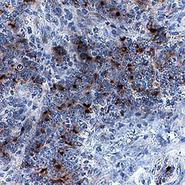
TIMP-1 was detected in immersion fixed paraffin-embedded sections of human colon cancer tissue using Goat Anti-Human TIMP-1 Antigen Affinity-purified Polyclonal Antibody (Catalog # AF970) at 1 µg/mL for 1 hour at room temperature followed by incubation with the Anti-Goat IgG VisUCyte™ HRP Polymer Antibody (Catalog # VC004). Before incubation with the primary antibody, tissue was subjected to heat-induced epitope retrieval using Antigen Retrieval Reagent-Basic (Catalog # CTS013). Tissue was stained using DAB (brown) and counterstained with hematoxylin (blue). Specific staining was localized to cytoplasm and extracellular space. View our protocol for IHC Staining with VisUCyte HRP Polymer Detection Reagents.Applications for Human TIMP-1 Antibody
Immunohistochemistry
Sample: Immersion fixed paraffin-embedded sections of human colon cancer tissue
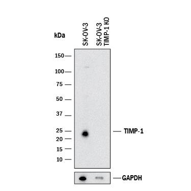
Knockout Validated
Simple Western
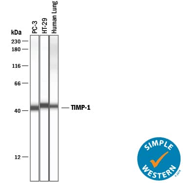
Sample: PC‑3 human prostate cancer cell line, HT‑29 human colon adenocarcinoma cell line, and human lung tissue
Western Blot
Sample: Human lung tissue, human prostate tissue, PC‑3 human prostate cancer cell line, HT‑29 human colon adenocarcinoma cell line, and SK‑OV‑3 human ovarian adenocarcinoma cell line
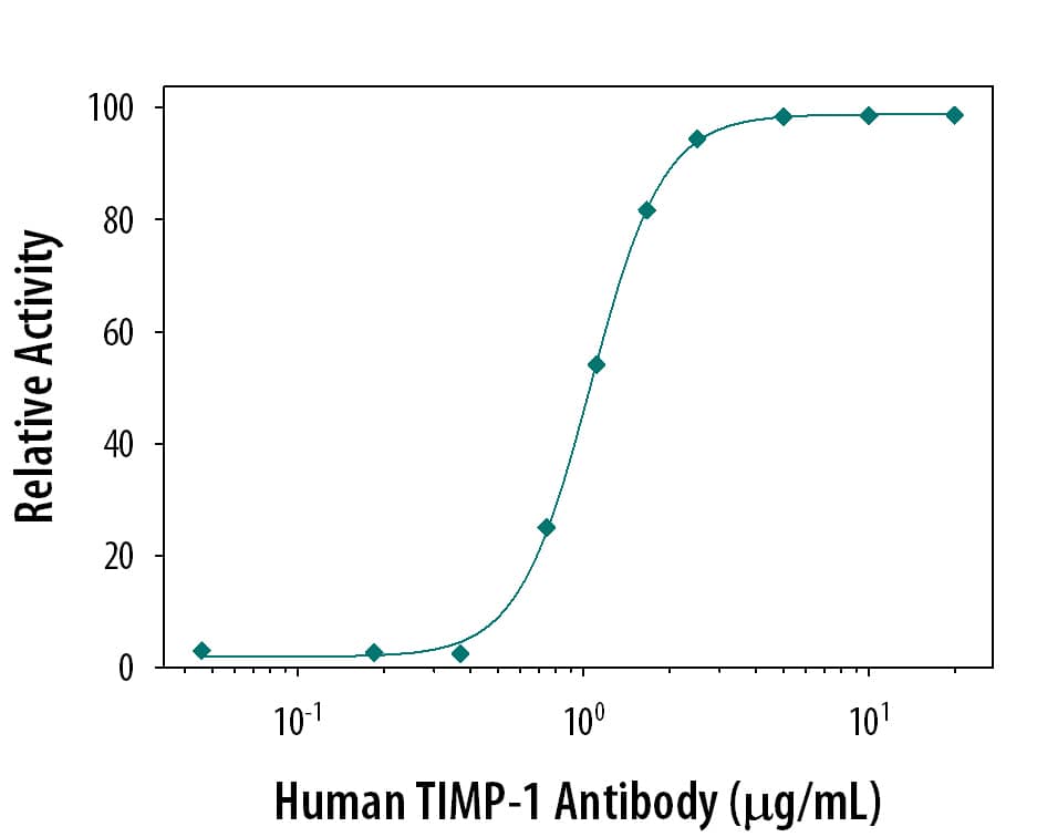
Neutralization
Reviewed Applications
Read 3 reviews rated 4.3 using AF970 in the following applications:
Formulation, Preparation, and Storage
Purification
Reconstitution
Formulation
Shipping
Stability & Storage
- 12 months from date of receipt, -20 to -70 °C as supplied.
- 1 month, 2 to 8 °C under sterile conditions after reconstitution.
- 6 months, -20 to -70 °C under sterile conditions after reconstitution.
Background: TIMP-1
Tissue inhibitors of metalloproteinases or TIMPs are a family of proteins that regulate the activation and proteolytic activity of the zinc enzymes known as matrix metalloproteinases (MMPs). There are four members of the family, TIMP-1, TIMP-2, TIMP-3 and TIMP-4. TIMP-1 is a glycoprotein with a molecular mass of 28 kDa produced by a wide range of cell types. TIMP-1 inhibits active MMP-mediated proteolysis by forming an N-terminal, non-covalent binary complex with the MMP active site. TIMP-1 also associates C-terminally with Pro-MMP-9 in a complex which may play a role in regulating activation. Independent of MMPs, TIMP-1 has been shown to have a role in tissue homeostasis.
Long Name
Alternate Names
Gene Symbol
UniProt
Additional TIMP-1 Products
Product Documents for Human TIMP-1 Antibody
Product Specific Notices for Human TIMP-1 Antibody
For research use only
