Human TRA-1-85/CD147 PE-conjugated Antibody
R&D Systems, part of Bio-Techne | Catalog # FAB3195P


Conjugate
Catalog #
Key Product Details
Species Reactivity
Validated:
Human
Cited:
Human
Applications
Validated:
Flow Cytometry
Cited:
Flow Cytometry, Immunocytochemistry, Immunohistochemistry
Label
Phycoerythrin (Excitation = 488 nm, Emission = 565-605 nm)
Antibody Source
Monoclonal Mouse IgG1 Clone # TRA-1-85
Product Specifications
Immunogen
2120Ep human embryonal carcinoma cell line
Specificity
Detects human TRA‑1‑85 antigen in flow cytometry.
Clonality
Monoclonal
Host
Mouse
Isotype
IgG1
Scientific Data Images for Human TRA-1-85/CD147 PE-conjugated Antibody
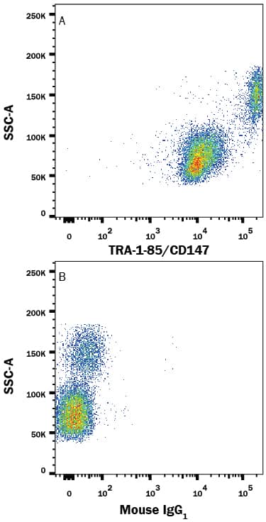
Detection of TRA-1-85/CD147 in Human PBMCs by Flow Cytometry.
Human peripheral blood lymphocytes and monocytes were stained with either (A) Mouse Anti-Human TRA-1-85/CD147 PE-conjugated Monoclonal Antibody (Catalog # FAB3195P) or (B) Mouse IgG1Phycoerythrin Isotype Control (Catalog # IC002P). View our protocol for Staining Membrane-associated Proteins.Detection of Mouse TRA-1-85/CD147 by Flow Cytometry
Flow cytometry analysis of total cell fractions of dissociated cells from PDX models.A) Basal-like xenograft cells. B) Luminal-like xenograft cells. A and B displays pseudo-color dot plots (left panels) and histograms (right panels). Freshly harvested xenografts were minced and the whole cell suspensions were washed and stained with monoclonal antibody towards EpCAM, TRA-1-85 (filled blue in histograms), H2-kd (red line in lower histogram) and Hoecst-33342 (intensity measure for DNA content of cells, grey contours in both histograms. Left peak indicate mouse cells, right peak indicate human cells). The population positive for both EpCAM and TRA-1-85, i.e the human tumor cells, are indicated with a circle in the dot plots. C) Flow cytometry analysis of double stained samples (marker of interest and EpCAM/Tra-1-85) of the Luminal-like PDX model. Flow cytometry histograms show the distribution of the markers indicated in the figure. Filled blue histogram represents EpCAM positive tumor cell population, and the EpCAM negative population (mouse stroma cells) is indicated by the red line. Grey contour represent unstained control. Image collected and cropped by CiteAb from the following publication (https://pubmed.ncbi.nlm.nih.gov/25419568), licensed under a CC-BY license. Not internally tested by R&D Systems.Detection of Mouse TRA-1-85/CD147 by Flow Cytometry
Flow cytometry analysis of total cell fractions of dissociated cells from PDX models.A) Basal-like xenograft cells. B) Luminal-like xenograft cells. A and B displays pseudo-color dot plots (left panels) and histograms (right panels). Freshly harvested xenografts were minced and the whole cell suspensions were washed and stained with monoclonal antibody towards EpCAM, TRA-1-85 (filled blue in histograms), H2-kd (red line in lower histogram) and Hoecst-33342 (intensity measure for DNA content of cells, grey contours in both histograms. Left peak indicate mouse cells, right peak indicate human cells). The population positive for both EpCAM and TRA-1-85, i.e the human tumor cells, are indicated with a circle in the dot plots. C) Flow cytometry analysis of double stained samples (marker of interest and EpCAM/Tra-1-85) of the Luminal-like PDX model. Flow cytometry histograms show the distribution of the markers indicated in the figure. Filled blue histogram represents EpCAM positive tumor cell population, and the EpCAM negative population (mouse stroma cells) is indicated by the red line. Grey contour represent unstained control. Image collected and cropped by CiteAb from the following publication (https://pubmed.ncbi.nlm.nih.gov/25419568), licensed under a CC-BY license. Not internally tested by R&D Systems.Applications for Human TRA-1-85/CD147 PE-conjugated Antibody
Application
Recommended Usage
Flow Cytometry
10 µL/106 cells
Sample: Human peripheral blood lymphocytes and monocytes
Sample: Human peripheral blood lymphocytes and monocytes
Formulation, Preparation, and Storage
Purification
Protein A or G purified from hybridoma culture supernatant
Formulation
Supplied in a saline solution containing BSA and Sodium Azide.
Shipping
The product is shipped with polar packs. Upon receipt, store it immediately at the temperature recommended below.
Stability & Storage
Protect from light. Do not freeze.
- 12 months from date of receipt, 2 to 8 °C as supplied.
Background: TRA-1-85/CD147
References
- Williams, B.P. et al. (1988) Immunogenetics. 27:322.
- Spring, F.A. et al. (1997) Eur. J. Immunol. 27:891.
Alternate Names
OKa Blood Group Antigen, TRA185
Entrez Gene IDs
682 (Human)
Gene Symbol
BSG
Additional TRA-1-85/CD147 Products
Product Documents for Human TRA-1-85/CD147 PE-conjugated Antibody
Product Specific Notices for Human TRA-1-85/CD147 PE-conjugated Antibody
For research use only
Loading...
Loading...
Loading...
Loading...
Loading...


