Human TRAIL R3/TNFRSF10C Antibody
R&D Systems, part of Bio-Techne | Catalog # MAB6301

Key Product Details
Validated by
Species Reactivity
Validated:
Cited:
Applications
Validated:
Cited:
Label
Antibody Source
Product Specifications
Immunogen
Ala26-Ala221
Accession # O14798
Specificity
Clonality
Host
Isotype
Scientific Data Images for Human TRAIL R3/TNFRSF10C Antibody
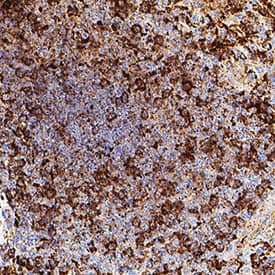
Detection of TRAIL R3/TNFRSF10C in Human Spleen.
TRAIL R3/TNFRSF10C was detected in immersion fixed paraffin-embedded sections of human spleen using Mouse Anti-Human TRAIL R3/TNFRSF10C Monoclonal Antibody (Catalog # MAB6301) at 15 µg/ml for 1 hour at room temperature followed by incubation with the Anti-Mouse IgG VisUCyte™ HRP Polymer Antibody (Catalog # VC001) or the HRP-conjugated Anti-Mouse IgG Secondary Antibody (Catalog # HAF007). Before incubation with the primary antibody, tissue was subjected to heat-induced epitope retrieval using VisUCyte Antigen Retrieval Reagent-Basic (Catalog # VCTS021). Tissue was stained using DAB (brown) and counterstained with hematoxylin (blue). Specific staining was localized to the membrane. View our protocol for IHC Staining with VisUCyte HRP Polymer Detection Reagents.Detection of Human TRAILR3/TNFRSF10C by Flow Cytometry
Flow cytometry studies of scFv-Fc-scTRAIL molecules and Fc-scTRAIL on Colo205 and HCT116 cells(A) Expression levels of EGFR, HER2, HER3, EpCAM, and TRAIL-receptors were investigated after treatment of the cells with medium or 250 ng/ml BZB for 16 h. (B) Serial dilutions of the molecules were analyzed. (C) 20 nM scTRAIL units were analyzed after preincubation of the cells with PBA or a 200-fold molar excess of the respective blocking antibody. Binding of Fc-scTRAIL was measured in the presence of all blocking antibodies separately and is here represented as the mean of blocking with all different antibodies. Bound molecules were detected via anti-FLAG-PE. relative MFI, relative median fluorescence intensity. Pairwise comparisons were performed by unpaired t test (two-tailed; *P < 0.05; **P < 0.01; ***P < 0.001; ns, P > 0.05). Image collected and cropped by CiteAb from the following publication (https://www.oncotarget.com/lookup/doi/10.18632/oncotarget.24379), licensed under a CC-BY license. Not internally tested by R&D Systems.Applications for Human TRAIL R3/TNFRSF10C Antibody
Immunohistochemistry
Sample: Immersion fixed paraffin-embedded sections of human spleen
Western Blot
Sample: Recombinant Human TRAIL R3/TNFRSF10C Fc Chimera (Catalog # 630-TR)
Human TRAIL R3/TNFRSF10C Sandwich Immunoassay
Formulation, Preparation, and Storage
Purification
Reconstitution
Formulation
*Small pack size (-SP) is supplied either lyophilized or as a 0.2 µm filtered solution in PBS.
Shipping
Stability & Storage
- 12 months from date of receipt, -20 to -70 °C as supplied.
- 1 month, 2 to 8 °C under sterile conditions after reconstitution.
- 6 months, -20 to -70 °C under sterile conditions after reconstitution.
Background: TRAIL R3/TNFRSF10C
Human TRAIL R3, also called DcR1 (decoy receptor 1), LIT, and TRID and designated TNFRSF10C, is a glycosyl-phosphatidylinositol-linked membrane protein which binds TRAIL (Apo2 Ligand) with high affinity. TRAIL R3 has the TRAIL-binding extracellular cysteine-rich domains but lacks the intracellular signalling domain. As a result, binding of TRAIL to TRAIL R3 does not transduce an apoptosis signal. Expression of TRAIL R3 has been shown to protect cells bearing TRAIL R1 and/or TRAIL R2 from TRAIL-induced apoptosis. A second TRAIL decoy receptor, which binds TRAIL with high-affinity but antagonizes TRAIL-induced apoptosis, named TRAIL R4, DcR2 or TRUNDD, has also been reported. The human soluble TRAIL R3/Fc chimera neutralizes the ability of TRAIL to induce apoptosis.
References
- Sheridan, J.P. et al. (1997) Science 277:818.
- Golstein, P. (1997) Curr. Biol. 7:R750.
Long Name
Alternate Names
Entrez Gene IDs
Gene Symbol
UniProt
Additional TRAIL R3/TNFRSF10C Products
Product Documents for Human TRAIL R3/TNFRSF10C Antibody
Product Specific Notices for Human TRAIL R3/TNFRSF10C Antibody
For research use only

